Posts

Oral Leukoplakia

Image
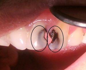
Oral leukoplakia  (OL) is a clinical term for a nonremovable white lesion that is not easily recognisable as any particular condition and therefore requires further investigation. Oral leukoplakia manifests as patches that are bright white and sharply defined. The surfaces of the patches are slightly raised above the surrounding mucosa. Oral leukoplakia may be homogenous (uniform lesion often with a fissured surface), or nonhomogeneous (with surface irregularity and textural or colour variation for example speckled-see below given photograph.

Stages and Phases of Anaesthesia

Assessment of the depth of anaesthesia  Anaesthesia has been described as a series of four Stages.  Stage 1 The period between administration of an anaesthetic and loss of consciousness.  Stage 2 The period after loss of consciousness, which may include actions such as uncontrolled movement, delirium, vocalization. Stage 3  The level at which surgery can be performed. Stage 3 anaesthesia is divided into four planes .  Plane 1 : "light" anaesthesia - the animal still has blink and swallowing reflexes, and regular respiration. Plane 2 : "surgical" anaesthesia - the animal has lost blink reflexes, pupils become fixed and respiration is regular. Plane 3 : "deep" anaesthesia - the animal starts losing the ability to use the respiratory muscles and breathing becomes shallow; may require assisted ventilation. The surgeries are performed in this plane. Plane 4 : the animal loses all respiratory effort, and breathing may stop entirely.  Stage 4 Anaesthetic cr...

Pulpitis

Image
Photograph showing Pulpal hyperaemia. While bacteria are still some distance from the pulp, acid permeating along the dentinal tubules gives rise to dilation of the blood vessels, oedema and a light cellular inflammatory infiltrate in the pulp [1] What is Pulpitis? Pulpitis is the inflammation of the pulp. It is the most common cause of pain in young persons. Types It is of two types. Reversible Irreversible Irreversible pulpitis has been divided into further two types Acute pulpitis Chronic pulpitis Causes of Pulpitis

Root Apex-Anatomical And Physiological Foramen

Image
Anatomical and physiological forament of the root   Apical constriction (Physiological foramen) is conside­­red narrowest diameter of the root canal located at the cement-dentinal junction. it is the apical limit of root canal preparation preparation. it is also known as the histological foramen, because it is located at the junction between the pulpal connective and interstitial loose connective tissues of the periodontal ligament. not visible on x-ray (we may find it only using WL determination methods).

Dental Caries-Part 6: Prevention

Image
Fig1: Caries free beautiful teeth with proper maintenance # The carious process can be termed as metabolic activity in the plaque. The result of the metabolic activity may not be anything to see or there may be demineralisation resulting in a visible carious lesion. As we have discussed earlier, plaque is the cause of caries and a tooth completely free of plaque will not have caries. However, it is not always possible to show a strong association between the presence of dental plaque and caries. Mostly because people are unable to completely remove plaque themselves, even when guided properly. In addition, although the bacterial biofilm is the cause of caries, there are other factors involved. This is the reason that caries is described as a multifactorial disease. These factors, when combined, may increase or decrease the rate of demineralization. 

Dental Caries-Part 5: Clinical Aspect of Caries Pathology

Image
Arrested caries and remineralization         Pre-Cavitation, or “white spot” caries lesions, can stop when the balance between demineralization and remineralization is changed in favor of remineralization. This could follow the restriction of sucrose, the application of fluoride, or the loss of an adjacent tooth to a proximal caries. This loss of tooth uncovers the area of stagnation and allows proper oral hygiene procedures. The source of the calcium and phosphate for remineralization of the lesion is saliva and plaque. The caries progresses slowly, and even under natural conditions, about 50% of proximal enamel lesions may show no radiographic evidence of progression for 3 years, showing that a small change may be needed to encourage reversal of the process. Although remineralization can bring the mineral content of an enamel lesion closer to that of the original enamel, the deposition is irregular and disorganized at the single crystal level, and the structure of ...

Dental Caries-Part 4: Dentine Caries

Image
Normal Structure of Dentin Dentine is the part of the tooth that is covered by enamel and covers the pulp tissues. It comprises of inorganic hydroxyapatite 45-50%, organic matrix 30% and water 25%. It is a vital tissue as opposed to enamel. It is elastic and permeable. Therefore, it is never possible for a restorative material to create a totally hermetic seal between itself and the cavity wall. Dentine consists of dentinal tubules, peritubular dentine and inter tubular dentine. Dentinal tubules contain extensions of the odontoblast cells that line along the inner surface of the dentine and remain in the pulp. The peritubular dentine makes the walls of the dentinal tubules, are devoid of collagen matrix and are highly mineralised. The inter tubular dentine is the main structural component; present between the dentinal tubules; consists of hydroxyapatite crystals lying in the collagen matrix. 

Dental Caries-Part 3: Enamel Caries

Image
  Clinical and Histological Features of Dental Caries Now we shall describes the clinical features of carious lesions on smooth, occlusal, and root surfaces.  We shall relate the clinical features to their histological features. We shall consider Enamel and Dentine together, the reasons being: As a clinician, you will see them in the same way. You can not understand changes in dentine during caries progression and caries arrest without considering the spread of the enamel lesion. Changes in Dentine occur before the enamel lesion cavitates. Removal of the biofilm will arrest the lesion in dentine as well as the lesion in enamel. The lesion, in both enamel and dentine, entirely reflects the activity of the bacterial biofilm. Before I start talking about the clinical and histological features of dental caries, You must know the  Basic Structure of Enamel Sound enamel consists of crystals of hydroxyapatite packed tightly together in an orderly arrangement which is known as en...

Dental Caries-Part 3: Video series

Image
Lecture Series on Dental Caries To watch video lectures, click at the following links 1. Introduction 2. Etiology of dental caries 3. Bacterial plaque in dental caries 4. Microbiology of dental caries 5. Sucrose and dental caries 6. Acid production in dental plaque 7. Saliva and dental caries 8. Early enamel caries

Dental Caries-Part 2: Treatment Planning

Image
In this chapter, you will know about the treatment planning of dental caries . In previous chapter you studies the etio-pathogenesis and clinical characteristics of dental caries . If you have not gone through it, it is advised that you study the previous chapter before proceeding further. Mechanism of Remineralisation of Enamel When the oral environment of a person is favourable where the pH is above 5.5 and saliva contains enough calcium and phosphate ions, the remineralisation process of enamel occurs. The supersaturated saliva acts as driving force for remineralisation. In a non cavitated enamel caries lesion, the original crystalline structure of rods remains intact. When it is etched, it acts as nucleating agent for remineralisation . When trace amount of fluoride ions is added to the environment, it enhances the remineralisation process by enhancing the precipitation of calcium and phosphate. The inclusion of fluoride ions results in the formation of fluorapatite crystals in e...

Dental Caries-Part 1: Etio-pathogenesis and Clinical Features

Image
This lesson presents basic definitions, terminologies, etiologies, demineralisation-remineralisation of enamel and clinical characteristics of the caries lesion in the context of clinical  operative dentistry.   Definitions of Dental Caries and Dental Plaque   Dental caries is defined as a multifactorial, transmissible, infectious oral disease caused primarily by the complex interaction of cariogenic oral flora (biofilm) with fermentable dietary carbohydrates on the tooth surface over time. Dental plaque is a gelatinous mass of bacteria adhering to the tooth surface. Carious lesions occur only under the plaque. The plaque bacteria metabolises the refined carbohydrate (sucrose mainly) for energy production and produces organic acids as a by product. These acids cause dissolution of crystalline structures of enamel that result in caries lesions of the tooth.  

Seven Ways to Prevent Dental Caries in Your Child

Image
Dental caries is an endemic disease in children. It is caused by a bacterial infection. The causative bacteria are known as Streptococcus Mutans and Lactobacilli. Although the dental caries is caused by bacteria, the initiation and progression of dental caries depend on several factors. The less the risk factors present, the less are the chances of dental caries development in your child’s tooth. I shall discuss those factors for you so that you can understand the developmental process of dental caries and take appropriate measures to prevent developing it in your son’s or daughter’s mouth. Broadly, we can categorize a child belonging to either high risk or low risk category. The children of high-risk category have higher chances of developing dental caries compared to those of lower category. The rampant caries develops rapidly in young children and involves all anterior teeth within a very short period. Early intervention may prevent its development. So, my advice to you, visit a den...

Oral Malodour / Foetor / Bad Breath / Halitosis

Image
Halitosis is a common social condition that affects a considerable portion of the general population. The prevalence of halitosis is close to 50% in a few populations. Under few circumstances, this oral condition may cause embarrassment, depression and make relationships more difficult. Some studies have investigated whether the psychology of the patients might have some influence on the complaints about halitosis, or even on halitosis itself. In a study that included more than one thousand participants and self-reported halitosis, scientists have reported that poor oral hygiene and general anxiety are associated with halitosis. [1] Halitosis is divided into intraoral, extraoral, pseudohalitosis and halitophobia. Intraoral halitosis, responsible for 85% of the cases of halitosis, is subdivided into physiologic (genuine) halitosis or pathologic halitosis. Extraoral halitosis occurs when malodor appears with no oral cause, as in the case of pulmonary causes. Both patients with pseudohali...

Basic Reason of Fear in a Four Years Old Child Who is Aggressive in Dental Office

The quality of dental treatment of a child depends on one's cooperation and repo with a dentist in the treatment room. How a dentist manages a child depending upon the understanding the child's  behaviour  pattern.    There are certain guidelines that are useful in helping a child to show a positive & compliant  behaviour  while getting the dental treatment. These guidelines have been prepared from the findings and principles of  behavioural  dentistry,  behavioural  psychology, developmental  psychology,  and  paediatric  psychology.