Malocclusion

MALOCCLUSION

Proposed by Edward H. Angle in 1890, the Angle Classifications are based on the relationship of the buccal groove of the mandibular first permanent molar and the mesiobuccal cusp of the maxillary first permanent molar. This classification is considered to be one of the most commonly used as its easy to use.

Bleeding Disorder

 Bleeding disorders: dental considerations

Acquired or congenital bleeding disorders of dental treatment concern include haemophilia, von Willebrand disease, other factor deficiencies and thrombocytopenia. Some systemic conditions also interfere with haemostasis, such as kidney, liver and bone marrow disorders.

Patients with bleeding disorders should be managed in a specialist setting, with appropriate consultation with the patient’s specialist or multidisciplinary team.

Bleeding Disorders

  1. haemophilia

  2. von Willebrand disease
  3. Other factor deficiencies 

  4. thrombocytopenia

Causes

Bleeding disorder may be due to defect in platelet activation, function and contact activation.  It may also be due to defect in clotting proteins or antithrombin function. 

The commonest caused of bleeding disorder are 

  1. warfarin
  2. von Willebrand disease
  3. aspirin

Warfarin is the commonest anticoagulant that interferes by preventing the production of clotting factors by blocking vitamin K.

von Willebrand disease is the most common inherited bleeding disorder.

Aspirin is the less commonly used drug for pain and more used in long term for its impairing ability for platelet function. One tablet of aspirin renders the platelet non-functional for almost one week. It is to be noted that it rarely causes significant post-operative bleeding.


How to confirm bleeding disorder of a patient

The single most important evaluation part is an adequate history since screening tests for bleeding disorders do not always detect mild defects. A history suggestive of a bleeding tendency must be taken seriously. History of previous dental extraction or tonsillectomy provide a useful guide. Physical examination is also necessary and for conformation of diagnosis, laboratory test are needed.

An accurate diagnosis is essential for replacement therapy, if needed and enable other management protocols to be followed.

The dentist must place special emphasis on the following that are suggestive of bleeding disorder:

  1. deep haemorrhage into  muscles, joints or skin (bruising)-suggests a clotting defect
  2. bleeding from or into skin or mucosae-suggests purpura
  3. most congenital bleeding disorders of significance become apparent in childhood
  4. however, mild haemophilia might escape the history part until the adult life if the child and the parents have managed well and avoided the injuries. A mild haemophiliac might give a history of mild oozing from an extraction socket for 2-3 weeks despite all local measures such as pressure pack and suturing etc
  5. history of blood transfusion or hospitalization might clearly give the idea of bleeding disorder in the same as a history of bleeding in a blood relative
  6. history of drugs such as anticoagulant, certain herbal products, systemic disease, for example, cirrhosis, HIV and kidney diseases can also impair platelet function 

Congestive Heart Failure

Congestive Heart Failure

Congestive heart failure or heart failure occurs when the heart muscle weakens and doesn't pump enough blood as it should. When this happens, blood often backs up and fluid can build up in the lungs, causing shortness of breath.

Certain heart conditions, for example, coronary artery disease or high blood pressure, gradually leave the heart too weak or stiff to fill and pump blood properly. 

Signs and symptoms of congestive heart failure 

  • Shortness of breath with activity or when lying down
  • Fatigue and weakness
  • Swelling in the legs, ankles and feet
  • Rapid or irregular heartbeat
  • Reduced ability to exercise
  • Persistent cough or wheezing with white or pink blood-tinged mucus
  • Swelling of the belly area (abdomen)
  • Very rapid weight gain from fluid build-up
  • Nausea and lack of appetite
  • Difficulty concentrating or decreased alertness
  • Chest pain if heart failure is caused by a heart attack

Prophylactic antibiotic regimen for infective endocarditis in dental procedures

Antibiotic prophylaxis is recommended for invasive dental procedures that involve the manipulation of gingival tissue or periapical region or perforation of the mucosa when performed on high-risk individuals. Australian guidelines have provided a list of dental procedures that are likely to cause a high incidence of bacteraemia that always require prophylaxis. These are as follows:

  • Tooth extraction.
  • Periodontal surgery, subgingival scaling and root planning.
  • Replantation of avulsed teeth.
  • Other surgical procedures such as implant placement or apicoectomy.

Procedures that cause a moderate incidence of bacteraemia might be considered for prophylaxis if multiple procedures are being conducted, in cases where the procedure is prolonged, or in the setting of periodontal disease.

Antibiotic prophylaxis is not recommended for procedures with a low possibility of bacteraemia such as:

  • Local anaesthetic injections.
  • Dental X-rays.
  • Treatment of superficial caries.
  • Orthodontic appliance placement and adjustment.
  • Following shedding of deciduous teeth.
  • After lip or oral trauma.


Patient Selection

The current infective endocarditis/valvular heart disease guidelines [2] state that use of preventive antibiotics before certain dental procedures is reasonable for patients with:

  1. prosthetic cardiac valves, including transcatheter-implanted prostheses and homografts;
  2. prosthetic material used for cardiac valve repair, such as annuloplasty rings and chords;
  3. a history of infective endocarditis;
  4. a cardiac transplant with valve regurgitation due to a structurally abnormal valve;
  5. the following congenital (present from birth) heart disease:
  • unrepaired cyanotic congenital heart disease, including palliative shunts and conduits
  • any repaired congenital heart defect with residual shunts or valvular regurgitation at the site of or adjacent to the site of a prosthetic patch or a prosthetic device

AP for a dental procedure not suggested [3]

  • Implantable electronic devices such as a pacemaker or similar devices 
  • Septal defect closure devices when complete closure is achieved 
  • Peripheral vascular grafts and patches, including those used for hemodialysis 
  • Coronary artery stents or other vascular stents 
  • CNS ventriculoatrial shunts 
  • Vena cava filters 
  • Pledgets 


Paediatric Patients


Congenital heart disease can indicate that prescription of prophylactic antibiotics may be appropriate for children. It is important to note, however, that when antibiotic prophylaxis is called for due to congenital heart concerns, they should only be considered when the patient has:

  • Cyanotic congenital heart disease (birth defects with oxygen levels lower than normal), that has not been fully repaired, including children who have had a surgical shunts and conduits.
  • A congenital heart defect that's been completely repaired with prosthetic material or a device for the first six months after the repair procedure.
  • Repaired congenital heart disease with residual defects, such as persisting leaks or abnormal flow at or adjacent to a prosthetic patch or prosthetic device[2].

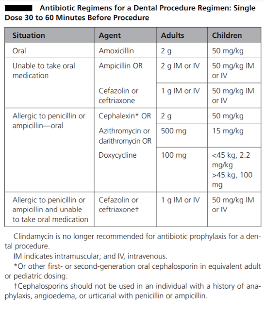
The prophylactic antibiotic should be effective against viridans group streptococci. The guidelines recommend 2 grams of amoxicillin given orally as a single dose 30-60 minutes before the procedure as the drug of choice for infective endocarditis prophylaxis. It has bactericidal activity against streptococci and enterococci. It reaches peak concentrations within one to two hours of oral administration, it has a short half-life of 1.5 hours, but therapeutic levels are maintained for nearly six hours. It has high oral bioavailability. The usual paediatric dosage is 50 mg/kg, with a maximum up to 2 gr. If the patient is unable to take oral medications, parenteral administration of 2 gr amoxicillin or ampicillin is considered as an alternative.

Oral or parenteral administration of cephalexin 2 gr for adults or 50 mg/kg for children, or parenteral administration of cefazolin or ceftriaxone 1 gr i.m./i.v. for adults or 50 mg/kg i.m./i.v for children are other alternatives. Cephalexin can be replaced by another first- or second-generation oral cephalosporin of equivalent dosage.

In patients hypersensitive to penicillin, guidelines are in agreement that the alternative drug of choice is clindamycin 600 mg (15-20 mg/kg up to 600 mg for children). It can be administered orally or intravenously 30-60 minutes before the procedure (as per European association of cardiologist [2], not as per ADA or AHA-see table). Clindamycin is a bacteriostatic protein synthesis inhibitor. Peak serum concentrations are achieved within 45 to 60 minutes after oral administration. Clindamycin is effective against streptococci and methicillin-sensitive staphylococci. However, some studies have questioned the potency of clindamycin prophylaxis. While ESC guidelines recommend solely clindamycin in penicillin-allergic patients, the AHA and Australian guidelines provide a variety of alternatives in this group of patients. The AHA guidelines recommend macrolides, 500 mg of azithromycin or clarithromycin (15 mg/kg for children). The Australian guidelines recommend glycopeptides; however, the ESC guidelines do not recommend glycopeptides and fluoroquinolones due to the lack of evidence on their efficacy. Cephalosporins should be refrained from use in patients who have encountered anaphylaxis, angioedema or urticaria related to penicillins.

It is important to administer prophylaxis before the procedure so that minimal inhibitory concentrations of the drugs will be present from the beginning of the procedure. If the patient cannot receive a prophylactic antibiotic before the procedure, it can be administered up to two hours after the procedure; however, delay in the treatment might lead to increased bacteraemia. If the patient needs multiple interventions, prophylaxis should be repeated with each. It is advised to finish necessary interventions in one or two sessions if possible. Given that consecutive exposures to the same antibiotic increase resistance rates, the healthcare provider might opt to choose different antibiotics for subsequent sessions. These might be the second-line alternative therapies mentioned in the guidelines or administering the patient a combination beta lactamase inhibitor such as amoxicillin-clavulanate or sulbactam-ampicillin. If the patient is already on antibiotic therapy with penicillins, the operation could be delayed until after the cessation of the antibiotic and restoration of the oral flora. If this is not possible, an alternative group of antibiotics could be preferred.


Ref:

1. https://www.escardio.org/Journals/E-Journal-of-Cardiology-Practice/Volume-16/vol16no33#:~:text=The%20prophylactic%20antibiotic%20should%20be,choice%20for%20infective%20endocarditis%20prophylaxis.

2. https://www.ada.org/resources/research/science-and-research-institute/oral-health-topics/antibiotic-prophylaxis

3.https://www.ahajournals.org/doi/pdf/10.1161/CIR.0000000000000969

Haemophilia

The laboratory findings in haemophilia will be as follows.

APTT (activated partial prothrombin time) -prolonged

PT (prothrombin time)-normal

BT (bleeding time)-normal [1] or increased [2]

Factor VIII-C- low

Factor VIIIR:Ag [von Willebrand factor] and factor VIIIR:RCo [Ristocetin cofactor]-normal

Ref:

  1. Crispian Scully, Roderick A. Cawson Medical problems in dentistry page 142 5th Ed. 
  2. https://www.cdc.gov/ncbddd/hemophilia/diagnosis.html


Oral Erythroplakia

Oral Erythroplakia


Erythroplakia is a clinical term for a potentially malignant fiery red lesion that cannot be attributed to any particular condition.


Signs and Symptoms


Lesions are usually asymptomatic and isolated, and commonly appear on the floor of the mouth, tongue, soft palate and buccal mucosa. Lesions may appear as smooth, velvety, granular or nodular plaques, often with clear margins.

Antimicrobials used in dentistry

Antimicrobials used in dentistry

Chemotherapy is the use of chemicals to destroy or inhibit the growth of cells. Two broad classes of chemotherapeutic agents are used in pharmacology: 

  1. antimicrobials and 
  2. anticancer drugs. 

The basis of antimicrobial chemotherapy is a differential sensitivity of the patient and microbe cells to the action of the drug. The drug may affect a structural component of the target cell which is not found in the patient, for example, the bacterial cell wall. Alternatively, a chemotherapeutic agent may inhibit a metabolic pathway peculiar to the microbe cells, for example, synthesis of folate.

Parotid Fistula

     Normally there is one opening of the parotid gland which is located in buccal vestibule opposite the upper 2nd molar tooth.

     Parotid fistula is a patent tract connecting a parotid gland or duct to the exterior apart from the parotid duct opening.

Photo 1. Pre-operative picture of parotid fistula with leakage of serous fluid from the fistulous tract and scarring of surrounding area (red circle) [1]

     Parotid fistula may be of two types

    1. Glandular: It arises directly from gland. It shows minimal discharge during rest or eating.
2. Ductal: It arises from duct. It shows profuse discharge during eating.

Parotid fistula may be extra oral or intraoral.

Extraoral fistulas are seen in the preauricular region or near the angle of mandible (see photo 1 and 2).

Photo 2. showing discharge of serous fluid from the right cheek in the angle of mandible region [2]


Causes
1. After superficial parotidectomy.
2. After drainage of parotid abscess.
3. After biopsy or Trauma.
4. Post surgical

Clinical Features
1.  Discharging fistula in the parotid region of the face, and discharge is more during eating.
2. Tenderness and induration.
3. Trismus if it gets infected

     Diagnosis
1.  Sialography to find out the origin of the fistula whether from the parotid gland or duct or ductules.
2. Fistulogram or CT fistulogram.
3. Culture of discharge if infection is suspected
4. MRI to assess soft tissues involvement

    Treatment
Ø Surgical stripping of the fistula tract
Ø Anticholinergics in post-operative period- Hyoscine bromide (Probanthine) reduce discharge
Ø Immediate post surgical fistulas can close spontaneously in such cases
Ø Newman Seabrock's operation: used for removal of anomalous arotid fistula
Ø If there is stenosis at the orifice of the Stenson's duct, papillotomy at the orifice may help.
Ø Total conservative parotidectomy is done in failed cases conserving the facial nerve
 
Ref:

Injections Techniques

Darsogluteal Intramuscular Injections


Kaposi sarcoma


Kaposi sarcoma (in AIDS):

Important points to remember about Kaposi Sarcoma

Kaposi's sarcoma is a type of cancer that forms in the lining of blood and lymph vessels.

Kaposi's sarcoma or oral cavity

Kaposi's sarcoma of the skin


Clinical Features

  • It is the most common malignancy in AIDS.
  • It is associated with the infection with a virus called the Kaposi sarcoma associated herpesvirus (KSHV), also known as human herpesvirus 8 (HHV8).
  • The commonly affected sites are palate, gingiva, tongue, and oropharynx or the skin of the face and feet.
  • It is seen early in the course of the disease. It can sometimes be confused with Bacillary angiomatosis.
  • There is a specific histologic stain for Kaposi sarcoma known as Warthin-Starry stain.
  • With the use of HAART, incidence of KS is decreasing and soon NHL may become the most common malignancy associated with AIDS.


Cemento Osseous Dysplasia

Cemento-osseous dysplasia (COD) is a benign fibro-osseous lesion of bone characterized by the replacement of normal bone by fibrous tissue and subsequently followed by its calcification with osseous and cementum-like material. It arises from the fibroblasts of the periodontal ligaments.

It is mostly asymptomatic in nature and requires no treatment. When secondarily infected, it becomes symptomatic and intervention is required.
Orthopantamogram showing a well-defined radiopaque mass in the right mandible region extending from the distal root of 45 to the mesial root of 47 [1]


As per WHO, there are three clinical presentation of cemento-osseous dysplasia.

  1. Periapical
  2. Focal
  3. Florid

Periapical cemento-osseous dysplasia

These occur in the anterior mandible and involve only a few adjacent teeth.

Focal cemento-osseous dysplasia.

involve few teeth in posterior mandible

Florid cemento-osseous dysplasia or Familial gigantiform cementoma

It is a more extensive form. it occurs bilaterally in mandible or in all jaw quadrants.
Ref:

Periapical Granuloma

The cells of periapical granuloma which are predominantly lymphocytes increase by division at the periphery. 

There are hyperaemia and oedema of the PDL; localised increase in the vascularity leads to local bone resorption mediated by osteoclast mediated delayed hypersensitivity. In the specimen slide, cholesterol crystals having needle-like appearance, and eosinic hyaline bodies known as Rushton bodies are seen. Macrophages and multinucleated giant cells are also seen. Epithelium is present.

The cells in the centre are separated from their source of nutrition; hence degenerate and liquefy. This results in an epithelium lined cavity filled with fluid known as periapical cyst.

Treatment involves RCT with apicoectomy or extraction with curettage.

Sequelae of Infection of Dental Pulp

Periapical infection with Streptococci & Staphylococci

Majority of streptococci produce hyaluronidase, an enzyme that dissolves hyaluronic acid which is a universal intercellular cementing substance. It helps in the spread of infection. Usually staphylococci are good producers of hyaluronidase, so there is no spread of infection and the infection becomes localised in the form of abscess in case of staph infection.

Oral Cancer

Oral cancer is associated with significant morbidity and mortality. Early presentations of oral cancer are usually asymptomatic, whereas late presentations include pain, discomfort, reduced mobility of the tongue, increased mobility of the teeth or an inability to wear dentures. Oral cancer varies in appearance and can mimic many other oral mucosal diseases.

Squamous cell carcinoma of the left anterior ventral surface of the tongue
Squamous cell carcinoma of the left mandibular alveolus

Oral cancer can mimic many other oral mucosal diseases, so early specialist referral is required for investigation and biopsy of any suspicious lesion. 

Any suspicious lesion needs early specialist referral for investigation and biopsy.

Squamous cell carcinoma is the most common oral malignancy, which arises from the epithelium of the oral cavity. Oral squamous cell carcinoma can affect any part of the oral mucosa; however, it most commonly occurs on the lateral surfaces of the tongue, the floor of the mouth or the gingivae. 


Risk factors for oral squamous cell carcinoma  

  1. advanced age 
  2. male gender 
  3. smoking or tobacco use 
  4. alcohol use 
  5. infection by oncogenic viruses (eg human papillomavirus) 
  6. personal or family history of squamous cell carcinoma of the head and neck 
  7. history of cancer therapy 
  8. prolonged immunosuppression 
  9. areca nut (betel quid) chewing. 
  10. Genetic susceptibility, environment, occupation and diet may also contribute to the development of oral squamous cell carcinoma. 

Cancers originating from the salivary glands and supporting nonepithelial tissues are less common than squamous cell carcinoma. Metastatic cancers to the oral soft tissues and jawbones commonly originate from primary malignancies in the breast, prostate, kidneys or lungs. Leukaemia and lymphoma may also present in the oral cavity. 


The treating specialist should perform the biopsy of an oral mucosal lesion. In rural or remote areas where a delay in specialist review is expected, seek expert advice on biopsy technique. A punch biopsy is not appropriate.


References:

  1. Therapeutic guidelines (Oral & Dental) 2019