Burning Mouth Syndrome

The diagnosis of burning mouth syndrome is by the process of exclusion. When we can not find the reason for burning mouth symptoms, the disease is diagnosed as burning mouth syndrome.

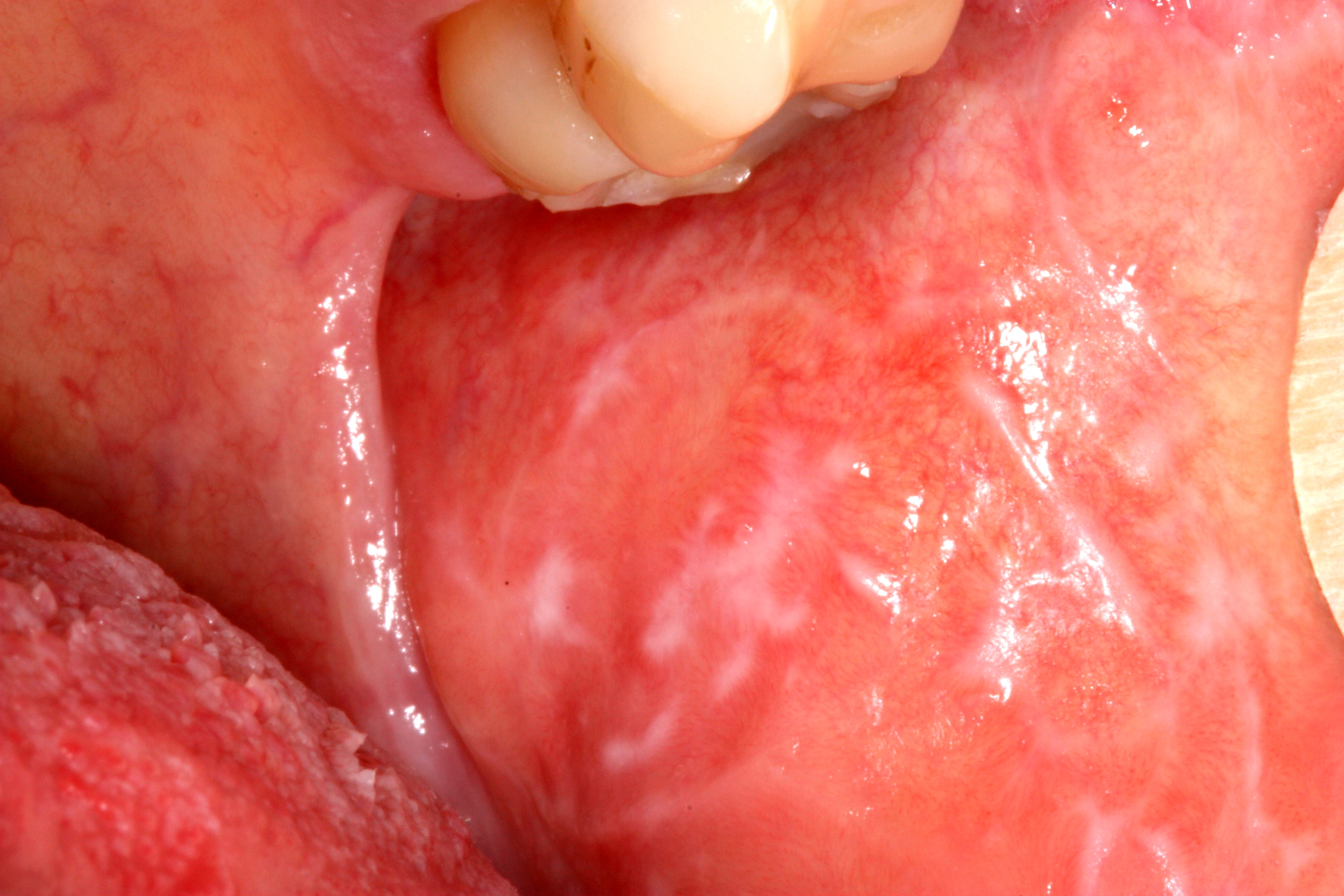
The characteristic symptom is a burning or scalding sensation of the tongue. Less frequently, we can find the coincident symptoms in hard palate and mucosal aspect of the lips. Additionally, patient may complain of a sensation of dry mouth with increased thirst, change in taste, such as a bitter or metallic taste or loss of taste. There may also be tingling, stinging or numbness in the mouth.

These symptoms can cause little inconvenience in mild cases. In severe cases, they can prevent patients from conducting normal daily activities. It has been found that in extreme cases, patients may show suicidal tendencies. 

In most cases, the burning sensation starts mild in the morning and increases in intensity as the day progresses. This type of presentation has the best prognosis.

You may find other signs and symptoms associated with burning mouth syndrome. These include:
  1. parafunctional habits, for example, unconsciously rubbing the tongue against the adjacent teeth and the hard palate which can cause traumatic abrasion of the filiform papillae on its dorsal surface
  2. dry mouth
  3. halitosis
  4. dysgeusia; most commonly a metallic taste

How will you diagnose and manage burning mouth syndrome

The diagnosis of burning mouth syndrome is done by the process of elimination. when you can not attribute the cause of the burning sensation in mucosa, the burning mouth syndrome diagnosis is made. Therefore, it is mandatory to know the diseases or conditions that produce burning mouth sensation. The common causes for burning mouth sensation are:

  1. local causes, for example, mucocutaneous conditions, fungal infections, rough dental surfaces
  2. systemic causes, for example, diabetes mellitus
  3. hypersensitivity to dental materials. Patients may say that he feels the problem is prosthesis-related. Hypersensitivity can be identified with skin patch testing, but rarely required
  4. drugs, for example, drugs that cause sensory neuropathy, taste aberrations or salivary gland hypofunction
You can use questionnaires to evaluate the impact of symptoms on the patient’s mood and quality of life.

You should understand that the management of burning mouth syndrome is complex. The most important part of the management is to make patient understand the condition. Try everything to make him understand that burning mouth syndrome is a chronic neuropathic pain syndrome; it doesn't matter, what's the trigger is.

Some patients may improve by discussion and counselling alone. Some may need lifestyle change and few will need pharmacological intervention.

Lifestyle changes to change the patient's response to external stressors. These include relaxation therapy, time management, exercise and community group discussion.

Pharmacological therapy includes psychotropic drugs, for example, tricyclic antidepressants, antiepileptics and clonazepam.

Most patient choose pharmacologic treatment for the management of burning mouth syndrome and it requires specialist intervention.


Ref: 
1. Therapeutic guidelines 2019


Oral Lichen Planus

Oral Lichen Planus on left mucosa [1]


Question: What is oral lichen planus?

Answer: It is a chronic inflammatory condition that affects the skin, nails, hair, and mucous membranes, characterised by purplish, itchy, flat eruptions.


Question: How common is the condition?

Answer: It is a common condition in India. Its cases are reported more than 10 lakh per year in India. 


Question: How much time does it need for recovery?

Answer: It can last several years or remains lifelong.


Question: Is the condition treatable? 

Answer: Treatments can help manage conditions. There is no known cure present. 


Question: Does diagnosis require lab tests or imaging? 

Answer: Its diagnosis rarely requires lab tests or imaging. 


Condition Highlights 

  1. It commonly occurs for ages 35-50. 
  2. It is more common in females. 
  3. Family history may increase likelihood to occur. 


Assessment of oral mucosal disease

Oral mucosal lesions are common. They can be due to physiological changes or a local disease. They may also be an oral manifestation of a skin condition, an adverse drug reaction or systemic disease, for example, gastrointestinal disease. To manage an oral mucosal disease successfully one requires an accurate diagnosis.

Now the question arises, how will we get an accurate diagnosis?

The correct answer is, by a thorough assessment of oral mucosa for a lesion.

Assessment for an oral mucosal lesion involves taking a full patient history. This includes a medication history too. Next we need to perform a thorough extraoral and intraoral examination and use diagnostic investigations where appropriate. One should have a high index of suspicion for oral cancer.

To recognise oral cancer one should be familiar with the risk factors for oral cancer. You can see the “Oral Cancer” topic to know about risk factors for oral cancer. You should also thoroughly know the red flag features of oral cancer. If any 'red flag' features are present or the diagnosis is not clear or the patient has not responded to initial treatment, early referral to an appropriate specialist is required.  An oral medicine specialist is the most appropriate specialist to diagnose and manage oral mucosal disease, but may not be accessible. Therefore,  an oral surgeon, dermatologist or otorhinolaryngologist are other options.

Failure to respond to initial treatment, an unclear diagnosis or the presence of any suspicious features could indicate malignancy and warrant early referral.

‘Red flag’ features of oral mucosal disease

  1. oral ulcers that have lasted for more than 2 weeks 

  2. orals ulcers that recur

  3. nontraumatic oral ulcers in children 

  4. pigmented lesions on the oral mucosa

  5. red, white or mixed red and white lesions on the oral mucosa of unknown origin or with features of potentially malignant disease, such as: induration, ulceration with rolled margins, fixation to underlying tissues, lesions in high-risk sites (eg lateral tongue, floor of mouth)

  6. facial or oral paraesthesia

  7. persistent oral mucosal discomfort with no obvious cause 

  8. lumps or swellings, including lymphadenopathy

  9. swelling, pain or blockage of a salivary gland, suggestive of salivary gland disease 

  10. suspected allergy or adverse reaction to dental materials for example oral lichenoid lesion, dry mouth that is not adequately relieved with artificial salivary products and nonpharmacological methods and dry mouth caused by systemic disease

  11. suspected oral manifestations of systemic disease, for example, syphilis, Behçet syndrome, HIV, inflammatory bowel disease, lichen planus, pemphigoid

  12. lesions occurring in immunocompromised patients, for example, patients with neutropenia or HIV infection

Some oral mucosal diseases are associated with significant morbidity and mortality, particularly oral potentially malignant disorders and oral cancer. Oral potentially malignant disorders include:

  1. oral leukoplakia 
  2. oral erythroplakia
  3. chronic hyperplastic candidiasis 
  4. actinic cheilitis
  5. oral lichen planus
  6. oral submucous fibrosis
  7. discoid lupus erythematosus 
  8. dyskeratosis congenita 
  9. epidermolysis bullosa

Oral potentially malignant disorders can become malignant at the site of the lesion, but also predict an increased risk of cancer at other sites in the mouth, even in clinically normal appearing oral mucosa.

The following conditions can be managed in general practice, provided there are no 'red flag' features present that would warrant referral:

  1. recurrent aphthous ulcerative disease

  2. traumatic oral ulcers

  3. oral candidiasis

  4. angular cheilitis

  5. oral mucocutaneous herpes simplex virus

  6. dry mouth

  7. oral mucositis

  8. amalgam tattoo

  9. geographic tongue

  10.  hairy tongue.

There are physiological causes of oral mucosal discolorations, for example, Fordyce spots which are ectopic sebaceous glands and leukoedema, that do not require active management.


Ref:

Therapeutic guidelines (oral & dental) 2019


Acute suppurative sialadenitis

Acute suppurative sialadenitis (including parotitis) is usually caused by Staphylococcus aureus. But sometimes it may be polymicrobial in adults. In acute suppurative sialadenitis, the glands are enlarged, often hot and tense, and pus may be expressed from the Stensen's duct. The patient is usually systemically unwell, dehydrated and has difficulty swallowing.

Intraoral view of purulence emanating from the parotid duct orifice in a patient with acute suppurative parotitis [1]. 


Management


Management of acute suppurative sialadenitis includes 
  • urgent referral to hospital for surgical review
  • rehydration 
  • culture and susceptibility testing of blood samples if the swelling is fluctuant, intraductal or surgical drainage; send pus for culture and susceptibility testing 
  • antibiotic therapy, given intravenously initially then orally once the patient can swallow. 

If S. aureus is identified in a blood culture, treat as S. aureus bacteraemia. If the results of blood culture indicate a polymicrobial bacteraemia, take expert advice. 


Intravenous antibiotic therapy for acute suppurative sialadenitis 


Initiate empirical antibiotic therapy for acute suppurative sialadenitis in conjunction with local intervention or drainage. Use flucloxacillin 2 g (in case of child: 50 mg/kg up to 2 g) intravenously, 6-hourly; afterwards you can switch to oral therapy once the patient can swallow.

For patients with risk factors for methicillin-resistant Staphylococcus aureus (MRSA) infection use vancomycin intravenously (for doses see details in iridium course); switch to oral therapy once the patient can swallow.

In some regions, based on local community acquired–MRSA susceptibility patterns, clindamycin or lincomycin is a suitable alternative to vancomycin.
  • clindamycin 600 mg (child: 15 mg/kg up to 600 mg) intravenously, 8-hourly; switch to oral therapy once the patient can swallow OR
  • lincomycin 600 mg (child: 15 mg/kg up to 600 mg) intravenously, 8-hourly; switch to oral therapy once the patient can swallow.

For patients with immediate nonsevere or delayed nonsevere hypersensitivity to penicillins and without risk factors for MRSA, use cefazolin 2 g (child: 50 mg/kg up to 2 g) intravenously, 8-hourly; switch to oral therapy once the patient can swallow.
 
For patients with immediate severe or delayed severe hypersensitivity to penicillins and without risk factors for MRSA, use clindamycin or lincomycin as above.


Oral continuation therapy for acute suppurative sialadenitis 


Initiate oral continuation therapy once the patient can swallow. If the results of culture and susceptibility
testing are available, modify oral therapy accordingly. If the results of susceptibility testing are not
available, use dicloxacillin 500 mg (child: 12.5 mg/kg up to 500 mg) orally, 6-hourly for a total of 10 days (oral & intravenous) or flucloxacillin 500 mg (child: 12.5 mg/kg up to 500 mg) orally, 6-hourly for a total of 10 days (intravenous plus oral).

For patients with risk factors for methicillin-resistant Staphylococcus aureus (MRSA) infection for whom results of susceptibility testing are not available, use trimethoprim+sulfamethoxazole 160+800 mg (child 1 month or older: 4+20 mg/kg up to 160+800 mg) orally, 12-hourly for a total of 10 days (intravenous plus oral) OR clindamycin 450 mg (child: 10 mg/kg up to 450 mg) orally, 8-hourly for a total of 10 days (intravenous plus oral)*

For patients with immediate nonsevere or delayed nonsevere hypersensitivity to penicillins and without risk factors for MRSA, use
cefalexin 500 mg (child: 12.5 mg/kg up to 500 mg) orally, 6-hourly.

For patients with immediate severe or delayed severe hypersensitivity to penicillins and without risk factors for MRSA, use trimethoprim+sulfamethoxazole or clindamycin as above.

*An oral liquid formulation of clindamycin is not commercially available; for formulation options for children or people with swallowing difficulties, see the Don’t Rush to Crush Handbook, published by the Society of Hospital Pharmacists of Australia.

Ref: 
  1. HealthJade
  2. Therapeutic Guidelines Australia 2019


Dental Burs

Diamond burs are generally used for reducing tooth structures to prepare cavities for restorations or place crowns or porcelain veneers. Diamonds may also be used to smooth, refine, and polish composite or porcelain material.

Tooth Whitening

Photograph showing before and and after teeth whitening procedure. 

Tooth whitening is done to correct the discoloration of a tooth. When we talk about discoloration of a tooth, we mean to say that  the color of a tooth has changed from its normal white color to  light yellow,  yellow,  light brown to dark brown  or from light grey to dark grey or  to complete black. The discolouration may be of one tooth or all the teeth may be discoloured. Discoloration of a tooth is caused by several factors. It may be due to deposition of external stains over the surfaces of a tooth or it may be because of the internal position of certain chemicals into the teeth structure at molecular level during the period when teeth were being formed in the mother’s womb.

Composites: Composition

Composites are tooth coloured restorative materials that are usually recommended for class III, IV and class I cavities with less or no occlusal stress and esthetics are important. Specially designed composites are used in almost 50% of class II restorations, although less durable in comparison to dental amalgam. Composites can be classified as microfilled, nanofilled, flowable, packable, all purpose and laboratory. Composites are used for provisional restorations and core build-ups and in fibre-reinforced posts.

Composites: Properties

You have read about the composition of dental composites in earlier class notes This article will speak about the properties. 

Properties of Composites

The important properties of the composites are as follows:
  1. Polymerisation shrinkage - should be low
  2. Water sorption - should be low
  3. Coefficient of thermal expansion - should be same as tooth
  4. Fracture resistance - should be high
  5. Wear resistance - should be high
  6. Radiopacity- should be high
  7. Bond strength to enamel & dentin - should be high
  8. Colour match to tooth structure - should be excellent
  9. Manipulation - should be easy
  10. Finishing and polishing - should be easy
Few of the above mentioned properties may be important for anterior than posteriors restorations and vice versa. The properties of 
microfilled and nanofilled composites are same while the microhybrid's differ from both of them.

Direct Esthetic Restorative Materials

Direct Esthetic Restorative Materials

There are four types of direct esthetic restorative materials currently in use. They are:

  1. Composites
  2. Compomers
  3. Hybrid Ionomers
  4. Glass Ionomers

Composites are dominating the materials used for direct esthetic restorations. Glass ionomers are primarily used for restorations of cervical eroded areas. Hybrid ionomers provide better esthetics than glass ionomers. Compomers provide improved handling and fluoride release when compared with composites.

Polysulfide Impression Materials

Permlastic is a polysulfide, condensation-cured, elastomeric impression material in three viscosities


Polysulfide impression materials are flexible but do not have the major changes in dimensions during storage like agar and alginate. Furthermore, the polysulfide impression is much stronger and more resistant to tearing than agar or alginate. It can be electroformed and therefore metal dies or models, in addition to gypsum models, can be prepared. 

Design of a Custom Tray for Final Impression for Complete Denture

The custom trays also known as special trays are fabricated so that the final impression of the patient’s edentulous mouth can be taken accurately with proper border extension. The selective pressure technique helps in making the impression in such a way that when the newly fabricated dentures are worn by a patient, they exert pressure only on those areas of the mouth that can withstand the masticatory load. The selective pressure technique of impression making spares the non-pressure bearing area of the edentulous jaw and thus eliminate the possibility of trauma resulting in pain and ulceration in the mucosa. The special tray for selective pressure technique is made on the primary cast that has been prepared by pouring the impression made in alginate or impression compound. The impression taken in impression compound records the mucosa in a compressed state whereas the impression taken with alginate records the mucosa in a non-compressed state. From these two opposite states of mucosa record, a dentist proceeds to prepare a special tray that is used to make an impression with selective pressure. We will learn here as to how to make a special tray on a cast that has been made from impression compound. 

Non-carious loss of tooth structure


Types, clinical features, Causes prevention & treatment


Non-carious loss of tooth structure is a problem that is often found in senior citizens and is a cause of many complaints. It is not a new entity but has acquired more attention in recent time.

Types of tooth wear

  1. Abrasion
  2. Attrition
  3. Erosion
  4. Demastication
  5. Abfraction

NEET 2022-23 Exam Dates Declared by NBE for NEET-MDS UG and PG

National Board of Examinations in Medical Sciences have declared the dates for NEET MDS, NEET PG & other exams. Candidates can check the exam dates below.

  1. NEET-MDS 2023: January 8, 2023
  2. DNB/DrNB Final Practical Examination – June 2022: October/November 2022
  3. Foreign Medical Graduate Examination (FMGE) December 2022, Foreign Dental Screening Test (FDST) 2022: December 4, 2022
  4. Formative Assessment Test (FAT) 2022: December 10, 2022
  5. DNB/DrNB Final Theory Examination – December 2022: December 21, 22, 23 and 24, 2022
  6. Fellowship Entrance Test (FET) 2022: January 20, 2023
  7. FNB Exit Examination 2022: February/March 2023
  8. DNB/DrNB Final Practical Examination – December 2022: Feb/March/April 2023
  9. NEET-PG 2023: March 5, 2023

Distribution of subject wise questions in NEET MDS examination.

The candidates are being advised to check the details and the updated information at the NBE website - https://natboard.edu.in/ as the dates mentioned are tentative and subject to approval and confirmation.


Harmful Effects of Excessive Radiation

The harmful effects of the excessive dose of the ionizing radiation can be divided into two types. These are as follows:  

  1. Deterministic effects and   
  2. Stochastic effects  

 We shall discuss them one by one in detail. 

Radiography: Radiation Safety

In earlier articles on radiography, you studied the effects of ionizing radiation on biological tissues. These effects can be divided into two types- Deterministic and Stochastic. Therefore, the radiation safety becomes of paramount importance white taking a radiograph.


Radiography: Radiation Physics

In this article termed as radiation physics, we shall talk about the ways, the x-rays are produced, the events that occur at atomic level during their production and how can one save oneself and others.

Production of X Rays  

Electrons that travel from the filament to the tungsten convert part of their kinetic energy into x-ray photons. This phenomenon occurs by the formation of bremsstrahlung and characteristic radiation.  

Radiography: Radiation Biology

Radiation Biology 

Radiation biology is the study of effects of ionizing radiation on living systems. In this article we shall talk about the harmful effects of ionizing radiation on tissues, how does it occur, and how sensitive different types of cells are?

The effects of radiation on the tissues can be divided into 2 broad categories. The Deterministic and Stochastic effects.


Cysts of the Jaws and Neck: Classification


Cysts of the Jaws and Neck 

Cysts can be classified in three types.

Odontogenic Cysts  

  1. Periapical (Radicular) Cyst  
  2. Lateral Periodontal Cyst  
  3. Gingival Cyst of the Newborn  
  4. Dentigerous Cyst  
  5. Eruption Cyst  
  6. Glandular Odontogenic Cyst  
  7. Odontogenic Keratocyst  
  8. Calcifying Odontogenic Cyst  

Nonodontogenic Cysts  

  1. Globulomaxillary Lesion  
  2. Nasolabial Cyst  
  3. Median Mandibular Cyst  
  4. Nasopalatine Canal Cyst  

Pseudocysts  

  1. Aneurysmal Bone Cyst  
  2. Traumatic (Simple) Bone Cyst  
  3. Static Bone Cyst (Stafne’s Bone Defect)  
  4. Focal Osteoporotic Bone Marrow Defect  
  5. Soft Tissue Cysts of the Neck  
  6. Branchial Cyst/ Cervical Lymphoepithelial Cyst  

Periapical/Radicular Cyst

A cyst is defined as “an epithelial lined pathologic cavity”. The periapical/radicular cyst is an odontogenic cyst. The classification of the cysts can be seen HERE. It is important to read for ADC Exams or NEET MDS purpose.

Periapical/Radicular cyst

Periapical cysts are inflammatory cysts. Their epithelial lining originates from the odontogenic epithelium of the tooth buds that remains within periodontal ligaments (epithelial rests of Malassez) after completion of tooth maturation. Due to inflammatory response, the epithelial rests of Malassez start proliferating and provide cystic lining.

Dentigerous/Follicular Cysts

Dentigerous (Follicular) Cysts are the second most commonly occurring odontogenic cysts after periapical cyst and the most common developmental cysts of the jaws. By definition, a dentigerous cyst is attached to the tooth cervix (enamel-cementum junction) and encloses the crown of the unerupted tooth.

Photograph1: Dentigerous cyst surrounding the crown of right mandibular third molar and going upward in ascending ramus. [1]

Etiology and Pathogenesis of Dentigerous Cyst 

A dentigerous cyst originates from the enamel organ remnant or reduced enamel epithelium. The expansion of the dentigerous cyst is related to epithelial proliferation, release of bone-resorbing factors, and an increase in cyst fluid osmolality. 

Clinical Features of Dentigerous Cyst 

Dentigerous cysts are most commonly seen associated with third molars and maxillary Canines. The peak incidence of dentigerous cysts occurs between twenty to 40 years. Males have more predilection with a ratio of l.6 to 1.  

Dentigerous cysts are generally symptomless. The delayed eruption is the most common indication of dentigerous cyst formation. This cyst can achieve significant size, occasionally causes cortical bone expansion but rarely reaches a size that predisposes the patient to a pathologic fracture. 

Radiographically, a dentigerous cyst manifests as a well-defined, unilocular or sometimes multilocular radiolucency with corticated margins in attached with the crown of an unerupted tooth. The concerned unerupted tooth is mostly displaced. In the mandible the related radiolucency may extend superiorly from the third molar site into the ramus or anteriorly and inferiorly along the body of the mandible. In maxillary dentigerous cysts in the canine region, extension into the maxillary sinus or to the orbital floor may be seen.

Histopathology.

The cyst is lined by stratified squamous epithelium. In a noninflamed dentigerous cyst the epithelial lining is nonkeratinized. It remains approximately four to six cell layers thick. Sometimes, numerous mucous cells, ciliated cells, and rarely, sebaceous cells may be found in the lining of the epithelium. The epithelium-connective tissue junction is generally flat. But when secondary inflammation established, epithelial hyperplasia may be noted.

Photomicrography 2: showed a thin non-keratinized epithelial lining composed of 2–3 layers of cuboidal epithelial cells and a fibrous connective-tissue wall loosely arranged. The arrow indicates an occasional mucous cell (bar = 0.2 mm) [1]

Differential diagnosis

When it is small, it is difficult to differentiate a dentigerous cyst from a large but normal dental follicle. When larger, the differential is essential that of lytic lesions of the jaw and includes:

  1. periapical cyst
  2. aneurysmal bone cyst
  3. ameloblastoma
  4. odontogenic keratocyst
  5. fibrous dysplasia
  6. Stafne cyst

Treatment

Removal of the associated tooth and enucleation of the soft tissue part is definitive therapy in most cases. When cysts affect significant portions of the mandible, exteriorization or marsupialization of the cyst is done to allow for decompression and subsequent shrinkage of the lesion followed by surgical enucleation. 

Ref:

  1. https://jcda.ca/article/c59
  2. https://radiopaedia.org/cases/dentigerous-cyst-9
  3. https://www.pathologyoutlines.com/topic/mandiblemaxilladentigerous.html
  4. https://www.nature.com/articles/modpathol2016191

Odontogenic Keratocyst

Odontogenic Keratocyst 


Odontogenic keratocyst (OKC) is a parakeratin lined cyst like lesion within bone.


OKCs mostly occur in the 2nd and 3rd decades of life; although can occur over a wide age range with male predilection. Odontogenic keratocyst comprises of 4 to 12 percent of all odontogenic cysts. Ninety percent of odontogenic keratocysts are solitary. Multiple cysts are found in Nevoid Basal Cell Carcinoma Syndrome / Gorlin Syndrome[1]. 


The most commonly involved body part in mandible, 65-85%; mostly posterior mandible. Normally, they are associated with third molars but may be present without associating with third molars; rarely occur in soft tissues.


They are considered to arise from dental lamina. 


Clinical Features 


Odontogenic keratocysts are often asymptomatic. They are incidentally discovered on radiographs. They can cause symptomatic swelling. They can cause parasthesia of lip and teeth. Symptoms of pain and drainage are found if secondarily infected. They can cause local bone and soft tissue destruction, but usually spare teeth and roots.


Swelling observed at the right buccal mucosa (dotted area) posterior to the orifice of Stensen’s duct-arrow head [3].


Odontogenic keratocyst-exposed anterior to the masseter muscle.


Radiographic features 


Mostly, they are seen as small unilocular radiolucent lesions with variable sclerotic margins. Larger lesions are often seen multilocular with variable scalloped margins. Therefore, They may resemble radicular cyst, dentigerous cyst, lateral periodontal cyst, and ameloblastoma.


Keratocystic Odontogenic tumer in orthopantomograph being shown as  multiple radiolucencies associated with mandibular anterior region, maxillary right and left as well as mandibular left impacted third molar tooth [5].

Classic look to a keratocyctic odontogenic tumor in the right mandible in the place of a former wisdom tooth. Unicystic lesion growing along the bone. Lesion was seen by oral surgeon on routine panoramic radiography without any symptoms [2].


Histology

Histological findings in the above slides are
a: The cyst wall is lined with parakeratinized squamous cells with a corrugated surface.
b: Nuclei of the cells in the basal layer are palisaded. The rete ridge of the epithelium is not evident [3]. 

Differential Diagnosis

  1. Ameloblastoma
  2. Dentigerous cyst
  3. Peri-apical cyst
  4. Lateral periodontal cyst

Prognosis

Recurrence rates of OKC are from 20% to 56% with enucleation alone. Resection have been reported to have no recurrences, but it may be considered excessive for a benign cyst.
Multiple lesions can occur when OKCs are associated with Nevoid Basal Cell Carcinoma Syndrome (NBCCS)/Gorlin Syndrome Therefore, early diagnosis and follow-up of the patient with OKC is important as there is always a possibility of developing other features of NBCCS in the future.

Treatment


Treatment is given taking into consideration of recurrence and morbidity. Following are the treatments given alone or in combination to a patient of odontogenic Keratocyst. 
  1. Decompression.
  2. Enucleation with possible curettage.
  3. Chemical curettage with Carnoy’s solution.
  4. Marsupialization.
  5. Resection.

Case Studies


For further studies on case reports, you can consult the reference number 4 and 5.


Ref:

  1. Morrison A. Odontogenic keratocyst. PathologyOutlines.com website. https://www.pathologyoutlines.com/topic/mandiblemaxillakeratocyst.html. Accessed September 11th, 2022.
  2. https://en.wikipedia.org/wiki/File:Classic_keratocystic_odontogenic_tumour.jpg
  3. https://www.ncbi.nlm.nih.gov/pmc/articles/PMC3837368/figure/F1/
  4. https://www.pathologyoutlines.com/caseofweek/case503.htm
  5. K. M. Veena, Rekha Rao, H. Jagadishchandra, Prasanna Kumar Rao, "Odontogenic Keratocyst Looks Can Be Deceptive, Causing Endodontic Misdiagnosis", Case Reports in Pathology, vol. 2011, Article ID 159501, 3 pages, 2011. https://doi.org/10.1155/2011/159501