Posts

Showing posts with the label dental caries

Dental Caries-Part 7: Mock Test Paper containing SBQ

Image
Dental Quiz INSTRUCTIONS: 1) The duration of the examination is 35 minutes. 2) After 20 minutes, it will show you a prompt for end of the exam duration. You will not be able to answer further questions. It will ask you to submit the answers. 3) Once you have finished answering the questions in time, press the submit button to see the answers. 4) After submission, you will see your result. The correct & wrong answers will be shown by a green tick and a red x in a circle respectively. You will get the correct answers in green colour and the explanations in magenta. 5)Your answers will not be saved automatically. Therefore, you need to write down your score yourself to keep a record of your progress 6) Once you are ready, press the start button to answer the questions. START Time Remaining: 0% Complete 1 2 3 4 5 6 7 8 9 1...
Dental Caries-Part 6: Prevention

Image
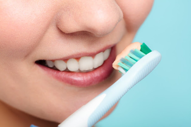
Fig1: Caries free beautiful teeth with proper maintenance # The carious process can be termed as metabolic activity in the plaque. The result of the metabolic activity may not be anything to see or there may be demineralisation resulting in a visible carious lesion. As we have discussed earlier, plaque is the cause of caries and a tooth completely free of plaque will not have caries. However, it is not always possible to show a strong association between the presence of dental plaque and caries. Mostly because people are unable to completely remove plaque themselves, even when guided properly. In addition, although the bacterial biofilm is the cause of caries, there are other factors involved. This is the reason that caries is described as a multifactorial disease. These factors, when combined, may increase or decrease the rate of demineralization. 

Dental Caries-Part 5: Clinical Aspect of Caries Pathology

Image
Arrested caries and remineralization         Pre-Cavitation, or “white spot” caries lesions, can stop when the balance between demineralization and remineralization is changed in favor of remineralization. This could follow the restriction of sucrose, the application of fluoride, or the loss of an adjacent tooth to a proximal caries. This loss of tooth uncovers the area of stagnation and allows proper oral hygiene procedures. The source of the calcium and phosphate for remineralization of the lesion is saliva and plaque. The caries progresses slowly, and even under natural conditions, about 50% of proximal enamel lesions may show no radiographic evidence of progression for 3 years, showing that a small change may be needed to encourage reversal of the process. Although remineralization can bring the mineral content of an enamel lesion closer to that of the original enamel, the deposition is irregular and disorganized at the single crystal level, and the structure of ...

Dental Caries-Part 4: Dentine Caries

Image
Normal Structure of Dentin Dentine is the part of the tooth that is covered by enamel and covers the pulp tissues. It comprises of inorganic hydroxyapatite 45-50%, organic matrix 30% and water 25%. It is a vital tissue as opposed to enamel. It is elastic and permeable. Therefore, it is never possible for a restorative material to create a totally hermetic seal between itself and the cavity wall. Dentine consists of dentinal tubules, peritubular dentine and inter tubular dentine. Dentinal tubules contain extensions of the odontoblast cells that line along the inner surface of the dentine and remain in the pulp. The peritubular dentine makes the walls of the dentinal tubules, are devoid of collagen matrix and are highly mineralised. The inter tubular dentine is the main structural component; present between the dentinal tubules; consists of hydroxyapatite crystals lying in the collagen matrix. 

Dental Caries-Part 3: Enamel Caries

Image
  Clinical and Histological Features of Dental Caries Now we shall describes the clinical features of carious lesions on smooth, occlusal, and root surfaces.  We shall relate the clinical features to their histological features. We shall consider Enamel and Dentine together, the reasons being: As a clinician, you will see them in the same way. You can not understand changes in dentine during caries progression and caries arrest without considering the spread of the enamel lesion. Changes in Dentine occur before the enamel lesion cavitates. Removal of the biofilm will arrest the lesion in dentine as well as the lesion in enamel. The lesion, in both enamel and dentine, entirely reflects the activity of the bacterial biofilm. Before I start talking about the clinical and histological features of dental caries, You must know the  Basic Structure of Enamel Sound enamel consists of crystals of hydroxyapatite packed tightly together in an orderly arrangement which is known as en...

Dental Caries-Part 3: Video series

Image
Lecture Series on Dental Caries To watch video lectures, click at the following links 1. Introduction 2. Etiology of dental caries 3. Bacterial plaque in dental caries 4. Microbiology of dental caries 5. Sucrose and dental caries 6. Acid production in dental plaque 7. Saliva and dental caries 8. Early enamel caries

Dental Caries-Part 2: Treatment Planning

Image
In this chapter, you will know about the treatment planning of dental caries . In previous chapter you studies the etio-pathogenesis and clinical characteristics of dental caries . If you have not gone through it, it is advised that you study the previous chapter before proceeding further. Mechanism of Remineralisation of Enamel When the oral environment of a person is favourable where the pH is above 5.5 and saliva contains enough calcium and phosphate ions, the remineralisation process of enamel occurs. The supersaturated saliva acts as driving force for remineralisation. In a non cavitated enamel caries lesion, the original crystalline structure of rods remains intact. When it is etched, it acts as nucleating agent for remineralisation . When trace amount of fluoride ions is added to the environment, it enhances the remineralisation process by enhancing the precipitation of calcium and phosphate. The inclusion of fluoride ions results in the formation of fluorapatite crystals in e...

Dental Caries-Part 1: Etio-pathogenesis and Clinical Features

Image
This lesson presents basic definitions, terminologies, etiologies, demineralisation-remineralisation of enamel and clinical characteristics of the caries lesion in the context of clinical  operative dentistry.   Definitions of Dental Caries and Dental Plaque   Dental caries is defined as a multifactorial, transmissible, infectious oral disease caused primarily by the complex interaction of cariogenic oral flora (biofilm) with fermentable dietary carbohydrates on the tooth surface over time. Dental plaque is a gelatinous mass of bacteria adhering to the tooth surface. Carious lesions occur only under the plaque. The plaque bacteria metabolises the refined carbohydrate (sucrose mainly) for energy production and produces organic acids as a by product. These acids cause dissolution of crystalline structures of enamel that result in caries lesions of the tooth.