Dens in Dente

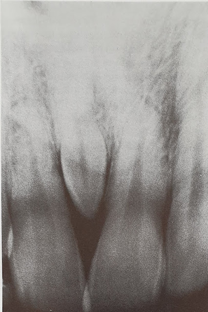
Dens invaginatus, dens in dente or tooth with in a tooth is a rare developmental anomaly. In it, the lingual pit is extended deep in to the crown or root, in later instance causing pulpitis. It is mostly seen in the maxillary lateral incisors but may be found in any anterior tooth.

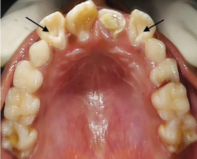
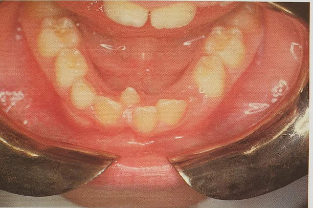
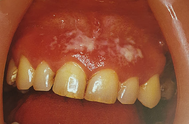
Dens in Dente-lateral incisors

The cause of dens in dente in not known but role of genetic factor can not be established. In early stage, it can be treated by filling or endodontic treatment, if pulp is involved.

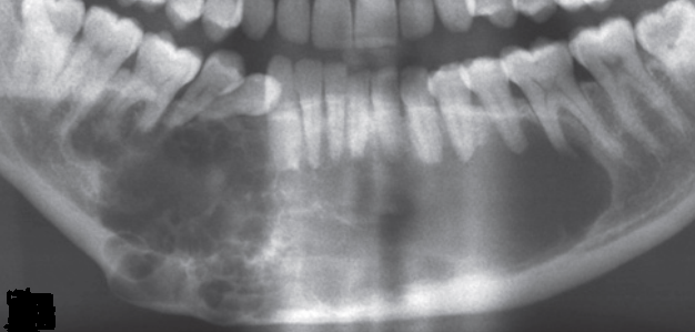
A. showing Type I Dens Invaginatus in lateral incisors. Note- periapical cyst. B. Type II dens invaginatus in second premolar and C. Type III dens invaginatus in mandibular canine. [1]

Ref: 
  1. https://www.omicsonline.org/scientific-reports/srep147.php

Hypertension

The 2017 ACC/AHA guideline for high BP in adults provides four BP categories based on the average of two or more in-office readings on 2 or more occasions:

  • Normal: Lower than 120 mm Hg systolic BP (SBP) and 80 mm Hg diastolic BP (DBP).
  • Elevated: 120–129 mm Hg SBP and lower than 80 mm Hg DBP.
  • Stage 1 hypertension: 130–139 mm Hg SBP or 80–89 mm Hg DBP.
  • Stage 2 hypertension: Higher than or equal to 140 mm Hg SBP or 90 mm Hg DBP.

Ameloblastoma

Ameloblastoma is a rare head and neck tumor with an estimated annual incidence of 0.5 per million population. They constitute 1% of tumors and cysts involving the jaws and accounts for approximately 10% of the odontogenic tumors. Ameloblastomas are originated from the epithelial lining of odontogenic cysts, enamel organ or dental lamina, stratified epithelium of oral cavity or displaced epithelial remnants. They are primarily seen in adults during the third and fourth decade of life with no gender preference and more frequently located in the mandible (80%), especially in the angle and ascending ramus [1].

Even though they are benign and slow-growing lesions, ameloblastomas exhibit locally destructive behavior with a high recurrence rate. Thus, most relapses (50% and even over 80%) occur during the first 5 years after the primary surgery. The major contributing factor for recurrence seems to be the inadequate initial surgical procedure rather than the histological type [1].

Radiographic Features

Radiographically, ameloblastoma typically forms round, cyst-like, radiolucent area with well-defined margins. The smallest lesions appear unilocular; whereas larger ameloblastoma may comprise a few large clustered cysts, giving 'soap-bubble’ or 'multilocular appearance' or  ‘honeycomb’ appearance (Fig 1) Expansion of the lesion may be on both, lingual and buccal side.
Fig. 1. Ameloblastoma: multilocular appearance


Differential Diagnosis

Other multilocular lesions that may mimic ameloblastoma radiologically include odontogenic keratocyst, giant-cell granuloma and odontogenic myxoma. Ameloblastoma with a single bony cavity simulate many types of cyst and tumour radiographically.

Treatment

The surgical options for ameloblastoma vary from simple enucleation (with or without bony curettage) to radical excision.


Ref: 
  1. Medina A, Velasco Martinez I, McIntyre B, Chandran R. Ameloblastoma: clinical presentation, multidisciplinary management and outcome. Case Reports Plast Surg Hand Surg. 2021;8(1):27-36. Published 2021 Feb 22. doi:10.1080/23320885.2021.1886854

Supernumerary Teeth

Extra numbers of teeth are known as supernumerary teeth. When they are present in the anterior maxilla in midline, they are known as mesiodens. When the extra teeth are present in the molar region as fourth molar, they are known as paramolar teeth. The anterior midline of the maxilla is the most common site whereas the maxillary molar area is the second most common site for supernumerary teeth.

Supernumerary tooth-mesiodens in anterior mandible in midline [1]


Radiograph showing mesiodens in anterior maxilla [1]


The investigation involves routine blood examination and IOPA or OPG radiographs. Depending on the anticipated level of difficulty of the surgery, additional investigations may be advised.

Treatment involves surgical extraction.


Ref:
  1. Oral pathology clinical pathologic correlation, Regezi, Sciubba, Jordan 4th Ed Saunders

Oral Candidiasis

Oral candidiasis is a fungal disease that is caused by Candida albicans. It looks like a white  or creamy plaque or patch that can be wiped off with the help of a cotton swab or a tooth brush leaving a red base.

Gingival thrush

It occurs due to disturbance in the oral microflora due to antibiotics, corticosteroid, Xerostomia , immune defects especially in HIV infection, immunosuppressant, leukaemia or lymphomas and diabetes. It rarely occurs in a healthy individuals except in neonates.

Chronic mucocutaneous candidosis: note the wide adherent plaque.

Gram stain smear shows the Candida albicans hyphae. It should be differentiated from Koplik's spot or Fordyce's granules.

The treatment involves treating the cause. Antifungal agents, for example, nystatin oral suspension or pastilles, amphotericin lozenges, or miconazole gel or tablets or fluconazole tablets can be given.


Ref:

1. Oral diseases 2nd Ed. Crispian Scully, Roderick A. Cawson Churchill Livingstone