Burning Mouth Syndrome

The diagnosis of burning mouth syndrome is by the process of exclusion. When we can not find the reason for burning mouth symptoms, the disease is diagnosed as burning mouth syndrome.

The characteristic symptom is a burning or scalding sensation of the tongue. Less frequently, we can find the coincident symptoms in hard palate and mucosal aspect of the lips. Additionally, patient may complain of a sensation of dry mouth with increased thirst, change in taste, such as a bitter or metallic taste or loss of taste. There may also be tingling, stinging or numbness in the mouth.

These symptoms can cause little inconvenience in mild cases. In severe cases, they can prevent patients from conducting normal daily activities. It has been found that in extreme cases, patients may show suicidal tendencies. 

In most cases, the burning sensation starts mild in the morning and increases in intensity as the day progresses. This type of presentation has the best prognosis.

You may find other signs and symptoms associated with burning mouth syndrome. These include:
  1. parafunctional habits, for example, unconsciously rubbing the tongue against the adjacent teeth and the hard palate which can cause traumatic abrasion of the filiform papillae on its dorsal surface
  2. dry mouth
  3. halitosis
  4. dysgeusia; most commonly a metallic taste

How will you diagnose and manage burning mouth syndrome

The diagnosis of burning mouth syndrome is done by the process of elimination. when you can not attribute the cause of the burning sensation in mucosa, the burning mouth syndrome diagnosis is made. Therefore, it is mandatory to know the diseases or conditions that produce burning mouth sensation. The common causes for burning mouth sensation are:

  1. local causes, for example, mucocutaneous conditions, fungal infections, rough dental surfaces
  2. systemic causes, for example, diabetes mellitus
  3. hypersensitivity to dental materials. Patients may say that he feels the problem is prosthesis-related. Hypersensitivity can be identified with skin patch testing, but rarely required
  4. drugs, for example, drugs that cause sensory neuropathy, taste aberrations or salivary gland hypofunction
You can use questionnaires to evaluate the impact of symptoms on the patient’s mood and quality of life.

You should understand that the management of burning mouth syndrome is complex. The most important part of the management is to make patient understand the condition. Try everything to make him understand that burning mouth syndrome is a chronic neuropathic pain syndrome; it doesn't matter, what's the trigger is.

Some patients may improve by discussion and counselling alone. Some may need lifestyle change and few will need pharmacological intervention.

Lifestyle changes to change the patient's response to external stressors. These include relaxation therapy, time management, exercise and community group discussion.

Pharmacological therapy includes psychotropic drugs, for example, tricyclic antidepressants, antiepileptics and clonazepam.

Most patient choose pharmacologic treatment for the management of burning mouth syndrome and it requires specialist intervention.


Ref: 
1. Therapeutic guidelines 2019


Oral Lichen Planus

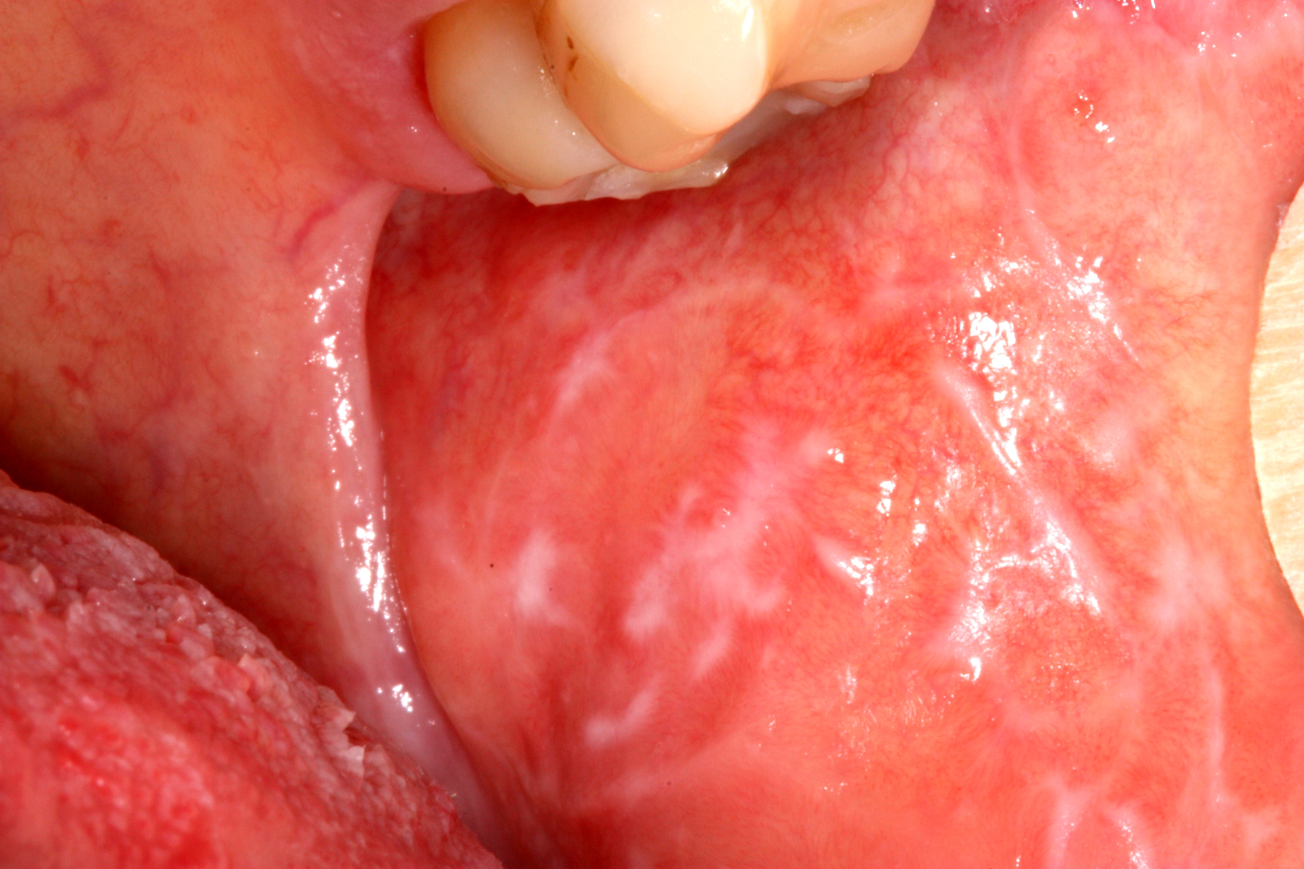
Oral Lichen Planus on left mucosa [1]


Question: What is oral lichen planus?

Answer: It is a chronic inflammatory condition that affects the skin, nails, hair, and mucous membranes, characterised by purplish, itchy, flat eruptions.


Question: How common is the condition?

Answer: It is a common condition in India. Its cases are reported more than 10 lakh per year in India. 


Question: How much time does it need for recovery?

Answer: It can last several years or remains lifelong.


Question: Is the condition treatable? 

Answer: Treatments can help manage conditions. There is no known cure present. 


Question: Does diagnosis require lab tests or imaging? 

Answer: Its diagnosis rarely requires lab tests or imaging. 


Condition Highlights 

  1. It commonly occurs for ages 35-50. 
  2. It is more common in females. 
  3. Family history may increase likelihood to occur. 


Assessment of oral mucosal disease

Oral mucosal lesions are common. They can be due to physiological changes or a local disease. They may also be an oral manifestation of a skin condition, an adverse drug reaction or systemic disease, for example, gastrointestinal disease. To manage an oral mucosal disease successfully one requires an accurate diagnosis.

Now the question arises, how will we get an accurate diagnosis?

The correct answer is, by a thorough assessment of oral mucosa for a lesion.

Assessment for an oral mucosal lesion involves taking a full patient history. This includes a medication history too. Next we need to perform a thorough extraoral and intraoral examination and use diagnostic investigations where appropriate. One should have a high index of suspicion for oral cancer.

To recognise oral cancer one should be familiar with the risk factors for oral cancer. You can see the “Oral Cancer” topic to know about risk factors for oral cancer. You should also thoroughly know the red flag features of oral cancer. If any 'red flag' features are present or the diagnosis is not clear or the patient has not responded to initial treatment, early referral to an appropriate specialist is required.  An oral medicine specialist is the most appropriate specialist to diagnose and manage oral mucosal disease, but may not be accessible. Therefore,  an oral surgeon, dermatologist or otorhinolaryngologist are other options.

Failure to respond to initial treatment, an unclear diagnosis or the presence of any suspicious features could indicate malignancy and warrant early referral.

‘Red flag’ features of oral mucosal disease

  1. oral ulcers that have lasted for more than 2 weeks 

  2. orals ulcers that recur

  3. nontraumatic oral ulcers in children 

  4. pigmented lesions on the oral mucosa

  5. red, white or mixed red and white lesions on the oral mucosa of unknown origin or with features of potentially malignant disease, such as: induration, ulceration with rolled margins, fixation to underlying tissues, lesions in high-risk sites (eg lateral tongue, floor of mouth)

  6. facial or oral paraesthesia

  7. persistent oral mucosal discomfort with no obvious cause 

  8. lumps or swellings, including lymphadenopathy

  9. swelling, pain or blockage of a salivary gland, suggestive of salivary gland disease 

  10. suspected allergy or adverse reaction to dental materials for example oral lichenoid lesion, dry mouth that is not adequately relieved with artificial salivary products and nonpharmacological methods and dry mouth caused by systemic disease

  11. suspected oral manifestations of systemic disease, for example, syphilis, Behçet syndrome, HIV, inflammatory bowel disease, lichen planus, pemphigoid

  12. lesions occurring in immunocompromised patients, for example, patients with neutropenia or HIV infection

Some oral mucosal diseases are associated with significant morbidity and mortality, particularly oral potentially malignant disorders and oral cancer. Oral potentially malignant disorders include:

  1. oral leukoplakia 
  2. oral erythroplakia
  3. chronic hyperplastic candidiasis 
  4. actinic cheilitis
  5. oral lichen planus
  6. oral submucous fibrosis
  7. discoid lupus erythematosus 
  8. dyskeratosis congenita 
  9. epidermolysis bullosa

Oral potentially malignant disorders can become malignant at the site of the lesion, but also predict an increased risk of cancer at other sites in the mouth, even in clinically normal appearing oral mucosa.

The following conditions can be managed in general practice, provided there are no 'red flag' features present that would warrant referral:

  1. recurrent aphthous ulcerative disease

  2. traumatic oral ulcers

  3. oral candidiasis

  4. angular cheilitis

  5. oral mucocutaneous herpes simplex virus

  6. dry mouth

  7. oral mucositis

  8. amalgam tattoo

  9. geographic tongue

  10.  hairy tongue.

There are physiological causes of oral mucosal discolorations, for example, Fordyce spots which are ectopic sebaceous glands and leukoedema, that do not require active management.


Ref:

Therapeutic guidelines (oral & dental) 2019


Acute suppurative sialadenitis

Acute suppurative sialadenitis (including parotitis) is usually caused by Staphylococcus aureus. But sometimes it may be polymicrobial in adults. In acute suppurative sialadenitis, the glands are enlarged, often hot and tense, and pus may be expressed from the Stensen's duct. The patient is usually systemically unwell, dehydrated and has difficulty swallowing.

Intraoral view of purulence emanating from the parotid duct orifice in a patient with acute suppurative parotitis [1]. 


Management


Management of acute suppurative sialadenitis includes 
  • urgent referral to hospital for surgical review
  • rehydration 
  • culture and susceptibility testing of blood samples if the swelling is fluctuant, intraductal or surgical drainage; send pus for culture and susceptibility testing 
  • antibiotic therapy, given intravenously initially then orally once the patient can swallow. 

If S. aureus is identified in a blood culture, treat as S. aureus bacteraemia. If the results of blood culture indicate a polymicrobial bacteraemia, take expert advice. 


Intravenous antibiotic therapy for acute suppurative sialadenitis 


Initiate empirical antibiotic therapy for acute suppurative sialadenitis in conjunction with local intervention or drainage. Use flucloxacillin 2 g (in case of child: 50 mg/kg up to 2 g) intravenously, 6-hourly; afterwards you can switch to oral therapy once the patient can swallow.

For patients with risk factors for methicillin-resistant Staphylococcus aureus (MRSA) infection use vancomycin intravenously (for doses see details in iridium course); switch to oral therapy once the patient can swallow.

In some regions, based on local community acquired–MRSA susceptibility patterns, clindamycin or lincomycin is a suitable alternative to vancomycin.
  • clindamycin 600 mg (child: 15 mg/kg up to 600 mg) intravenously, 8-hourly; switch to oral therapy once the patient can swallow OR
  • lincomycin 600 mg (child: 15 mg/kg up to 600 mg) intravenously, 8-hourly; switch to oral therapy once the patient can swallow.

For patients with immediate nonsevere or delayed nonsevere hypersensitivity to penicillins and without risk factors for MRSA, use cefazolin 2 g (child: 50 mg/kg up to 2 g) intravenously, 8-hourly; switch to oral therapy once the patient can swallow.
 
For patients with immediate severe or delayed severe hypersensitivity to penicillins and without risk factors for MRSA, use clindamycin or lincomycin as above.


Oral continuation therapy for acute suppurative sialadenitis 


Initiate oral continuation therapy once the patient can swallow. If the results of culture and susceptibility
testing are available, modify oral therapy accordingly. If the results of susceptibility testing are not
available, use dicloxacillin 500 mg (child: 12.5 mg/kg up to 500 mg) orally, 6-hourly for a total of 10 days (oral & intravenous) or flucloxacillin 500 mg (child: 12.5 mg/kg up to 500 mg) orally, 6-hourly for a total of 10 days (intravenous plus oral).

For patients with risk factors for methicillin-resistant Staphylococcus aureus (MRSA) infection for whom results of susceptibility testing are not available, use trimethoprim+sulfamethoxazole 160+800 mg (child 1 month or older: 4+20 mg/kg up to 160+800 mg) orally, 12-hourly for a total of 10 days (intravenous plus oral) OR clindamycin 450 mg (child: 10 mg/kg up to 450 mg) orally, 8-hourly for a total of 10 days (intravenous plus oral)*

For patients with immediate nonsevere or delayed nonsevere hypersensitivity to penicillins and without risk factors for MRSA, use
cefalexin 500 mg (child: 12.5 mg/kg up to 500 mg) orally, 6-hourly.

For patients with immediate severe or delayed severe hypersensitivity to penicillins and without risk factors for MRSA, use trimethoprim+sulfamethoxazole or clindamycin as above.

*An oral liquid formulation of clindamycin is not commercially available; for formulation options for children or people with swallowing difficulties, see the Don’t Rush to Crush Handbook, published by the Society of Hospital Pharmacists of Australia.

Ref: 
  1. HealthJade
  2. Therapeutic Guidelines Australia 2019