The main criterion for the division of keratinized epithelia is the presence or absence of nuclei in the keratinized layer viewed under light microscope. In the orthokeratinized epithelium the cell nuclei disappear in the keratinized layer, whereas in the parakeratinized epithelium flattened, highly condensed nuclei remain in the cell cytoplasm of the keratinized layer until cells exfoliate.
A coaching institution for ADC, ORE and Government dental job examinations.
Classification of Epithelia
INBDE - Integrated National Board Dental Examination
What is the INBDE?
The Integrated National Board Dental Examination (INBDE) is the 500-item licensing exam administered by the American Dental Association (ADA) which allows graduate dental students to practice in the United States of America. The content of the INBDE is formulated by the Joint Commission on National Dental Examinations (JCNDE).
An Introduction to Dental Crowns
Dental Crowns
Crowns are a far more complex procedure than most people realise. To make a good quality crown, a dentist has to work in within a fraction of a millimetre. There is almost no margin for error. Getting the appearance correctly and communicating accurately with the dental lab can be an issue. Added to these, discussions about the balance between appearance and maintaining tooth tissue can be tricky. It can take a lot of painstaking adjustment at every step. Add in time pressure and any other added difficulties and it's a recipe for a stressed dentist, particularly if the dentist has recently graduated with limited experience.
A calm and supportive dental nurse can work slickly with the dentist and makes the entire procedure easier. The roles of a good dental nurse and efficient lab technician are vital to the success of crowns.
Diagram showing transvers view of a tooth showing shape and thickness of a dental crown in pink colour.
- Planning for Making a Crown
The first stage is the treatment planning, and deciding whether or not to place a crown. A decision has to be made about whether a filling is sufficient, whether a crown is most appropriate, or whether the tooth has become unrestorable and an extraction is the only choice. Crowns can be quite destructive due to the amount of tooth material to be removed. On the contrary, the advantage of the crowns is that they can be protective to the tooth and can help to hold the tooth together just like a helmet on the head, preventing the tooth splitting outwards, specifically after a root canal treatment. No restorative material can replace original tooth tissue so unnecessary tooth destruction must be avoided. so although they certainly have their place, crowns should not be placed without good reason.
The next big question comes is whether the patient's priority is the appearance or saving tooth tissue. Sometimes choosing to aim for the optimum appearance can be done without endangering the pulp of the tooth but at other times a compromise has to be made in order to keep the tooth alive and healthy. Some patients will be willing to accept a metal-coloured tooth quite happily to keep as much of their own tooth as possible because the metal crowns can be made much thinner, but for others that will not be an acceptable option.
At most occasions, compromises are done and agreed upon, like a metal band around the gingival margin of the tooth, or just having the visible part of the crown tooth coloured and the rest metal coloured. Discussing the patients' priorities and making sure that they know what exactly they will get end of treatment is a very important part of the process, to ensure their happiness and satisfaction.
Another important part of the planing is checking whether the tooth is vital. Often crowns are placed following root canal treatment. Therefore, it is important to check that any infection under the tooth has improved. If the tooth is not root canal treated, then vitality tests and radiographic examination is a must for long term prognosis.
Crown Preparation
The first stage of the crown preparation appointment is normally making an impression for making a temporary crown while the permanent crown is being fabricated. The impression can be made using alginate or putty. Alginate needs to be mixed smoothly and the impression needs to be accurate with no big air bubble in the relevant area specially the margins. Putty needs to be used if the impression may be needed at a later date, as alginate impressions soon lose their shape.
Then comes the actual ‘crown preparation’ stage. Local anaesthetic is normally given to anaesthetise the tissues around the tooth being prepared. Cutting the tooth for the crown is the time where the dentist's real skill is needed. The amount of tooth tissue removed and the space needed for the crown depends on the material to be used for each part of the crown. A thicker porcelain is needed for porcelain whereas metal need to be kept thinner. For a patient, the difference between little tooth tissue removal and too much removal is fairly small. The dentist will try to create a clear margin, ideally finishing on natural tooth rather than filling material, for the dental lab to work on. On one hand, a written laboratory authorisation form will communicate a lot of the information about the crown to be made, the work done on the actual tooth can give a lot of information about what the dentist is intending for the tooth and how they want the lab to make the crown. The margin needs to be clear in order for it to be picked up clearly in the impression so that the lab has something useful to work with and so that the end result is good.
Shade Selection
After the crown prep is completed, the shade(s) for the crown needs to be decided on, in the case of porcelain. A shade guide is used and the dentist will look at different shades compared with the surrounding teeth. If the crown is full porcelain, rather than part porcelain with metal underneath, the dentist may also record the colour of the tooth underneath that is being crowned. This can then be communicated to the lab, to give them a greater idea of what they are working with, as the underlying colour can shine through and affect the colour of the crown needed. Any little details wanted, for example, stains or slight cracks on the tooth, can also be recorded and communicated.
Before the patient leaves, the tooth needs to be temporised. This is where the tooth is covered in order to keep it safe and healthy while the crown is made by the lab. It also helps to prevent movement or further eruption of the tooth that could prevent the crown fitting. The impression taken earlier in the process is used; temporary crown material is put into the impression and the impression is put back over the tooth. A small blob of the material is often put somewhere to give an indication of when the material is set. Once the material has set, the impression and temporary crown is removed. The edges are smoothed off using polishing discs. Temporary cement is then used for cementing the temporary crown in place.
The dental laboratory will then make the crown. This is also a very complex process, but largely falls outside of the clinical practice. The dental technician will fill the impression with dental stone to create a model/die of the tooth to work on. The lab will work to the instruction of the dentist, creating what has been asked for. The process will vary depending on whether it is a full metal, a porcelain fused to metal or a full porcelain crown.
Once the crown returns back from the lab, the dentist will check that it is as they expected. They will check if the crown goes on and off on the model perfectly.
Crown Cementation
The temporary crown will be removed, the temporary cement will be cleaned off the underlying tooth and a cement will be decided upon. The crown will be tried in and out so that the dentist is certain about the placement of the crown. Cement will then be mixed and put into the crown, the tooth will be dried, and the crown will be placed. Excess cement will be cleared away using floss and dental instruments. Pressure will be maintained on the crown so that the cement sets with the crown fully seated/in the correct position.
The patient will then be asked how it feels to bite on. Often it will be slightly ‘high’, so the dentist will mark the teeth using articulating paper, so that the heavy contacts show up. Adjustments will then be made so that the crown is comfortable and doesn't interfere with the patient's bite.
Crowns can be a fabulous restoration, helping to save teeth that would otherwise have to be extracted. They can also be a very aesthetic option, particularly those made of/with porcelain. Crowns can be a very complex and involved procedure, but really demonstrate the important roles that different members of the dental team play in producing an end result that really meets the needs of/pleases the patient.
Summary of stages:
- Deciding whether a crown is the most appropriate restoration
- Planning the material(s) of the crown
- Taking an impression to allow a temporary crown to be made
- Shaping the tooth ready for the crown
- Temporising the tooth
- The crown is made by the lab
- The temporary crown is replaced with the permanent crown
- Any necessary adjustments are made.
Bacterial Infection of the Oral Cavity
Bacterial lesions causing ulcerative conditions in the oral cavity
- Syphilis
- Gonorrhoea
- Tuberculosis
- Leprosy
- Actinomycosis
- Noma
Properties of Dental Materials- Terminology
You must know the properties of the materials used in dentistry to understand its function and how and why it works in oral cavity. It also helps in manipulating it. The main properties you should know about are following:
- Stress
- Strain
- Elasticity Modulus
- Elastic Limit
- Thermal Expansion
- Hygroscopic Expansion
- Setting Expansion
Dental Anatomy and Terminology
Anatomy of the tooth and surrounding tissues
Enamel: This is the hard, calcified substance that makes the surface of a crown of a tooth.
chamber of the tooth is surrounded by dentine.
Pulp: This is the organ at the centre of a tooth that contains blood vessels, connective and neural tissue, and cells that produce dentine-odontoblast. Blood vessels and neural tissue enter the tooth from the apex of the root.
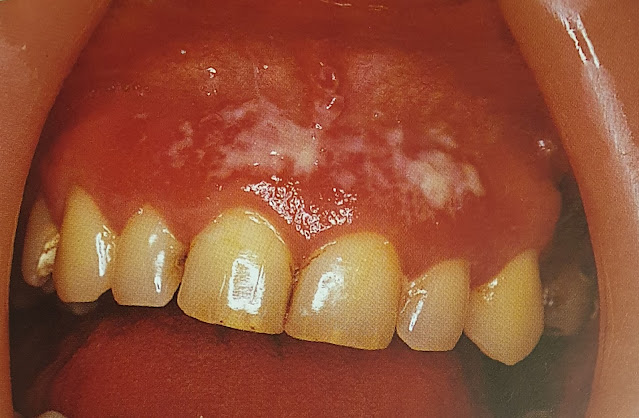
Periodontal ligament: This is the ligament that connects a tooth, by its root, to the supporting bone.
Cementum: This is the calcified tissue on the surface of the root of a tooth, which provides attachment for the periodontal ligament.
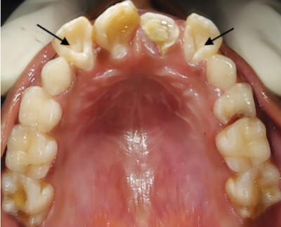
Fissure: It is a naturally occurring crevice in the enamel.
Crown: This is the part of the tooth that is visible and is above the gingival margin.
Root: This is the part of the tooth below the gingival margin; it is connected through cementum on its surface and the fibres of the periodontal ligament to the supporting bone.
Dental numbering system
There are numerous dental numbering systems to identify teeth and their
maturity. The most commonly used system in Australia is the Federation Dentaire
Internationale (FDI) system
The FDI numbering system divides the mouth into quadrants. The first
number indicates the quadrant and whether it is a primary or secondary tooth.
The second number indicates the tooth; tooth numbering begins at the central
incisor and counts backward to the molars.
Using the FDI numbering system, for adults, the quadrants are numbered as:
1. patient’s upper right is quadrant 1
2. patient’s upper left is quadrant 2
3. patient’s lower left is quadrant 3
4. patient’s lower right is quadrant 4
For primary teeth in children, the quadrants are numbered as:
1. patient’s upper right is quadrant 5
2. patient’s upper left is quadrant 6
3. patient’s lower left is quadrant 7
4. patient’s lower right is quadrant 8
Quick MCQ Test:
Face bow
![]() |
Face bow-for Whip mix Arcon articulator |
Face bow is a device that is used to transfer the relationship of upper jaw to the temporomandibular joint and the skull. The use of face bow are as follows:
- in full mouth rehabilitation
- when you want to give accurate crown and bridges
- when you need to alter VDO
- for diagnostic mounting
- for the correction of the occlusion
- when you want to use inter-occlusal records except when you are making tooth supported prosthesis or a single tooth restoration
- when you are using cusp form teeth
- when you need balanced occlusion
- when you want to do a gnathological study
![]() |
Face bow for Hanau Non-Arcon Articulator |
Multiple choice questions
INSTRUCTIONS:
Dens in Dente
Dens invaginatus, dens in dente or tooth with in a tooth is a rare developmental anomaly. In it, the lingual pit is extended deep in to the crown or root, in later instance causing pulpitis. It is mostly seen in the maxillary lateral incisors but may be found in any anterior tooth.
Dens in Dente-lateral incisors |
Hypertension
The 2017 ACC/AHA guideline for high BP in adults provides four BP categories based on the average of two or more in-office readings on 2 or more occasions:
- Normal: Lower than 120 mm Hg systolic BP (SBP) and 80 mm Hg diastolic BP (DBP).
- Elevated: 120–129 mm Hg SBP and lower than 80 mm Hg DBP.
- Stage 1 hypertension: 130–139 mm Hg SBP or 80–89 mm Hg DBP.
- Stage 2 hypertension: Higher than or equal to 140 mm Hg SBP or 90 mm Hg DBP.
Ameloblastoma
Ameloblastoma is a rare head and neck tumor with an estimated annual incidence of 0.5 per million population. They constitute 1% of tumors and cysts involving the jaws and accounts for approximately 10% of the odontogenic tumors. Ameloblastomas are originated from the epithelial lining of odontogenic cysts, enamel organ or dental lamina, stratified epithelium of oral cavity or displaced epithelial remnants. They are primarily seen in adults during the third and fourth decade of life with no gender preference and more frequently located in the mandible (80%), especially in the angle and ascending ramus [1].
Even though they are benign and slow-growing lesions, ameloblastomas exhibit locally destructive behavior with a high recurrence rate. Thus, most relapses (50% and even over 80%) occur during the first 5 years after the primary surgery. The major contributing factor for recurrence seems to be the inadequate initial surgical procedure rather than the histological type [1].
Radiographic Features
Fig. 1. Ameloblastoma: multilocular appearance |
Differential Diagnosis
Treatment
The surgical options for ameloblastoma vary from simple enucleation (with or without bony curettage) to radical excision.
- Medina A, Velasco Martinez I, McIntyre B, Chandran R. Ameloblastoma: clinical presentation, multidisciplinary management and outcome. Case Reports Plast Surg Hand Surg. 2021;8(1):27-36. Published 2021 Feb 22. doi:10.1080/23320885.2021.1886854
Supernumerary Teeth
Extra numbers of teeth are known as supernumerary teeth. When they are present in the anterior maxilla in midline, they are known as mesiodens. When the extra teeth are present in the molar region as fourth molar, they are known as paramolar teeth. The anterior midline of the maxilla is the most common site whereas the maxillary molar area is the second most common site for supernumerary teeth.
Supernumerary tooth-mesiodens in anterior mandible in midline [1]
Radiograph showing mesiodens in anterior maxilla [1]
The investigation involves routine blood examination and IOPA or OPG radiographs. Depending on the anticipated level of difficulty of the surgery, additional investigations may be advised.
Treatment involves surgical extraction.
- Oral pathology clinical pathologic correlation, Regezi, Sciubba, Jordan 4th Ed Saunders
Oral Candidiasis
Oral candidiasis is a fungal disease that is caused by Candida albicans. It looks like a white or creamy plaque or patch that can be wiped off with the help of a cotton swab or a tooth brush leaving a red base.
Gingival thrush |
It occurs due to disturbance in the oral microflora due to antibiotics, corticosteroid, Xerostomia , immune defects especially in HIV infection, immunosuppressant, leukaemia or lymphomas and diabetes. It rarely occurs in a healthy individuals except in neonates.
Chronic mucocutaneous candidosis: note the wide adherent plaque. |
Gram stain smear shows the Candida albicans hyphae. It should be differentiated from Koplik's spot or Fordyce's granules.
The treatment involves treating the cause. Antifungal agents, for example, nystatin oral suspension or pastilles, amphotericin lozenges, or miconazole gel or tablets or fluconazole tablets can be given.
Ref:
1. Oral diseases 2nd Ed. Crispian Scully, Roderick A. Cawson Churchill Livingstone
Malocclusion
MALOCCLUSION
Proposed by Edward H. Angle in 1890, the Angle Classifications are based on the relationship of the buccal groove of the mandibular first permanent molar and the mesiobuccal cusp of the maxillary first permanent molar. This classification is considered to be one of the most commonly used as its easy to use.