Pulpitis

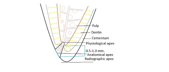
Photograph showing Pulpal hyperaemia. While bacteria are still some distance from the pulp, acid permeating along the dentinal tubules gives rise to dilation of the blood vessels, oedema and a light cellular inflammatory infiltrate in the pulp [1]

What is Pulpitis?

Pulpitis is the inflammation of the pulp. It is the most common cause of pain in young persons.


Types

It is of two types.

  1. Reversible

  2. Irreversible

Irreversible pulpitis has been divided into further two types

  1. Acute pulpitis

  2. Chronic pulpitis

Causes of Pulpitis

Root Apex-Anatomical And Physiological Foramen

Anatomical and physiological root apex
Anatomical and physiological forament of the root 

Apical constriction (Physiological foramen)

  • is conside­­red narrowest diameter of the root canal

  • located at the cement-dentinal junction.

  • it is the apical limit of root canal preparation preparation.

  • it is also known as the histological foramen, because it is located at the junction between the

  • pulpal connective and interstitial loose connective tissues of the periodontal ligament.

  • not visible on x-ray (we may find it only using WL determination methods).

Dental Caries-Part 6: Prevention

Fig1: Caries free beautiful teeth with proper maintenance #

The carious process can be termed as metabolic activity in the plaque. The result of the metabolic activity may not be anything to see or there may be demineralisation resulting in a visible carious lesion. As we have discussed earlier, plaque is the cause of caries and a tooth completely free of plaque will not have caries. However, it is not always possible to show a strong association between the presence of dental plaque and caries. Mostly because people are unable to completely remove plaque themselves, even when guided properly. In addition, although the bacterial biofilm is the cause of caries, there are other factors involved. This is the reason that caries is described as a multifactorial disease. These factors, when combined, may increase or decrease the rate of demineralization. 

Dental Caries-Part 5: Clinical Aspect of Caries Pathology

Arrested caries and remineralization  


Pre-Cavitation, or “white spot” caries lesions, can stop when the balance between demineralization and remineralization is changed in favor of remineralization. This could follow the restriction of sucrose, the application of fluoride, or the loss of an adjacent tooth to a proximal caries. This loss of tooth uncovers the area of stagnation and allows proper oral hygiene procedures. The source of the calcium and phosphate for remineralization of the lesion is saliva and plaque. The caries progresses slowly, and even under natural conditions, about 50% of proximal enamel lesions may show no radiographic evidence of progression for 3 years, showing that a small change may be needed to encourage reversal of the process. Although remineralization can bring the mineral content of an enamel lesion closer to that of the original enamel, the deposition is irregular and disorganized at the single crystal level, and the structure of the original enamel cannot be recovered. Despite this, remineralized lesions that have incorporated fluoride may be less prone to caries attack than intact enamel. Arrested enamel cavities can remain dull and white or more often discolored due to the incorporation of an extrinsic dye. This is known as inactive lesion or brown spot.   

Dental Caries-Part 4: Dentine Caries

Normal Structure of Dentin


Dentine is the part of the tooth that is covered by enamel and covers the pulp tissues. It comprises of inorganic hydroxyapatite 45-50%, organic matrix 30% and water 25%.
It is a vital tissue as opposed to enamel. It is elastic and permeable. Therefore, it is never possible for a restorative material to create a totally hermetic seal between itself and the cavity wall.
Dentine consists of dentinal tubules, peritubular dentine and inter tubular dentine. Dentinal tubules contain extensions of the odontoblast cells that line along the inner surface of the dentine and remain in the pulp. The peritubular dentine makes the walls of the dentinal tubules, are devoid of collagen matrix and are highly mineralised. The inter tubular dentine is the main structural component; present between the dentinal tubules; consists of hydroxyapatite crystals lying in the collagen matrix. 

Dental Caries-Part 3: Enamel Caries


Clinical and Histological Features of Dental Caries

Now we shall describes the clinical features of carious lesions on smooth,

occlusal, and root surfaces.  We shall relate the clinical features to their histological

features. We shall consider Enamel and Dentine together, the reasons being:

  1. As a clinician, you will see them in the same way.

  2. You can not understand changes in dentine during caries progression and caries arrest without considering the spread of the enamel lesion.

  3. Changes in Dentine occur before the enamel lesion cavitates. Removal of the biofilm will arrest the lesion in dentine as well as the lesion in enamel.

  4. The lesion, in both enamel and dentine, entirely reflects the activity of the bacterial biofilm.


Before I start talking about the clinical and histological features of dental caries, You must know the 

Basic Structure of Enamel


Sound enamel consists of crystals of hydroxyapatite packed tightly together in an orderly arrangement which is known as enamel prisms. The amount of hydroxyapatite ranges between 86 to 95%; the organic component between 1% to 2 % and water between 4% to 12% by volume. The total inorganic content of enamel ranges between 95% to 98% by mass, thats why it looks like crystals.


The crystals are so tightly packed that the enamel gets a glass-like appearance and appears translucent. This is the reason that it allows the varying degrees of yellow colour of the dentine to shine through it. Here, you should know that even though the crystal packing is very tight, each crystal is actually separated from its neighbours by tiny intercrystalline spaces or pores. These spaces are filled with water and organic material. When enamel is exposed to acids produced in the microbial biofilm, mineral is removed from the surface of the crystal which shrinks in size. Thus, the intercrystalline spaces enlarge and the tissue becomes more porous. This increase in porosity can be seen clinically as a white spot.

Dental Caries-Part 3: Video series


Lecture Series on Dental Caries

To watch video lectures, click at the following links

1. Introduction

2. Etiology of dental caries

3. Bacterial plaque in dental caries

4. Microbiology of dental caries

5. Sucrose and dental caries

6. Acid production in dental plaque

7. Saliva and dental caries

8. Early enamel caries


Dental Caries-Part 2: Treatment Planning

In this chapter, you will know about the treatment planning of dental caries. In previous chapter you studies the etio-pathogenesis and clinical characteristics of dental caries. If you have not gone through it, it is advised that you study the previous chapter before proceeding further.

Mechanism of Remineralisation of Enamel

When the oral environment of a person is favourable where the pH is above 5.5 and saliva contains enough calcium and phosphate ions, the remineralisation process of enamel occurs. The supersaturated saliva acts as driving force for remineralisation. In a non cavitated enamel caries lesion, the original crystalline structure of rods remains intact. When it is etched, it acts as nucleating agent for remineralisation. When trace amount of fluoride ions is added to the environment, it enhances the remineralisation process by enhancing the precipitation of calcium and phosphate. The inclusion of fluoride ions results in the formation of fluorapatite crystals in enamel rods which is more resistant to acid attack compared to calcium apatite of the natural enamel rods. Thus, the new enamel becomes resistant to caries process. 

Dental Caries-Part 1: Etio-pathogenesis and Clinical Features

This lesson presents basic definitions, terminologies, etiologies, demineralisation-remineralisation of enamel and clinical characteristics of the caries lesion in the context of clinical operative dentistry. 

Definitions of Dental Caries and Dental Plaque 

Dental caries is defined as a multifactorial, transmissible, infectious oral disease caused primarily by the complex interaction of cariogenic oral flora (biofilm) with fermentable dietary carbohydrates on the tooth surface over time.

Dental plaque is a gelatinous mass of bacteria adhering to the tooth surface. Carious lesions occur only under the plaque. The plaque bacteria metabolises the refined carbohydrate (sucrose mainly) for energy production and produces organic acids as a by product. These acids cause dissolution of crystalline structures of enamel that result in caries lesions of the tooth.  

Hepatitis B-Part 1: Epidemiology and Clinical Features

Overview 

Hepatitis is an inflammation of the liver that's caused by a variety of contagious viruses and non-infectious agents leading to a range of health problems, some of which can be fatal. There are five main strains of the hepatitis virus, known as types A, B, C, D and E. While their infection results in liver disease, they differ in important ways including modes of transmission, severity of the illness, geographical distribution and prevention methods. 

Particularly types B and C lead to chronic disease in hundreds of millions of people and together are the most common cause of liver cirrhosis, liver cancer and viral hepatitis related deaths. 

An estimated 354 million people worldwide having no access to testing and treatment live with hepatitis B or C. 

Some types of hepatitis are preventable through vaccination. A WHO study found that an estimated 4.5 million premature deaths could be averted in low-and middle- income countries by 2030 through vaccination, individual tests, drugs and education campaigns. WHO’s global hepatitis strategy, championed by all WHO Member States, aims to reduce new hepatitis infections by 90 and deaths by 65 between 2016 and 2030.


Hepatitis B is a potentially life-threatening liver infection caused by the hepatitis B virus (HBV). It can cause chronic infection and puts people at high risk of death from cirrhosis and liver cancer.

In this first part of the article, we shall discuss the following aspects of hepatitis:

  1. Epidemiology, 
  2. Mode of transmission
  3. Sign and symptoms 
  4. Groups at risk
  5. Relationship of HBV and HIV infection H
  6. How to confirm the diagnosis
  7. Sample MCQs