Cemento Osseous Dysplasia

Cemento-osseous dysplasia (COD) is a benign fibro-osseous lesion of bone characterized by the replacement of normal bone by fibrous tissue and subsequently followed by its calcification with osseous and cementum-like material. It arises from the fibroblasts of the periodontal ligaments.

It is mostly asymptomatic in nature and requires no treatment. When secondarily infected, it becomes symptomatic and intervention is required.
Orthopantamogram showing a well-defined radiopaque mass in the right mandible region extending from the distal root of 45 to the mesial root of 47 [1]


As per WHO, there are three clinical presentation of cemento-osseous dysplasia.

  1. Periapical
  2. Focal
  3. Florid

Periapical cemento-osseous dysplasia

These occur in the anterior mandible and involve only a few adjacent teeth.

Focal cemento-osseous dysplasia.

involve few teeth in posterior mandible

Florid cemento-osseous dysplasia or Familial gigantiform cementoma

It is a more extensive form. it occurs bilaterally in mandible or in all jaw quadrants.
Ref:

Periapical Granuloma

The cells of periapical granuloma which are predominantly lymphocytes increase by division at the periphery. 

There are hyperaemia and oedema of the PDL; localised increase in the vascularity leads to local bone resorption mediated by osteoclast mediated delayed hypersensitivity. In the specimen slide, cholesterol crystals having needle-like appearance, and eosinic hyaline bodies known as Rushton bodies are seen. Macrophages and multinucleated giant cells are also seen. Epithelium is present.

The cells in the centre are separated from their source of nutrition; hence degenerate and liquefy. This results in an epithelium lined cavity filled with fluid known as periapical cyst.

Treatment involves RCT with apicoectomy or extraction with curettage.

Sequelae of Infection of Dental Pulp

Periapical infection with Streptococci & Staphylococci

Majority of streptococci produce hyaluronidase, an enzyme that dissolves hyaluronic acid which is a universal intercellular cementing substance. It helps in the spread of infection. Usually staphylococci are good producers of hyaluronidase, so there is no spread of infection and the infection becomes localised in the form of abscess in case of staph infection.

Oral Cancer

Oral cancer is associated with significant morbidity and mortality. Early presentations of oral cancer are usually asymptomatic, whereas late presentations include pain, discomfort, reduced mobility of the tongue, increased mobility of the teeth or an inability to wear dentures. Oral cancer varies in appearance and can mimic many other oral mucosal diseases.

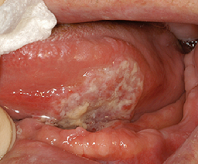
Squamous cell carcinoma of the left anterior ventral surface of the tongue
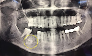
Squamous cell carcinoma of the left mandibular alveolus

Oral cancer can mimic many other oral mucosal diseases, so early specialist referral is required for investigation and biopsy of any suspicious lesion. 

Any suspicious lesion needs early specialist referral for investigation and biopsy.

Squamous cell carcinoma is the most common oral malignancy, which arises from the epithelium of the oral cavity. Oral squamous cell carcinoma can affect any part of the oral mucosa; however, it most commonly occurs on the lateral surfaces of the tongue, the floor of the mouth or the gingivae. 


Risk factors for oral squamous cell carcinoma  

  1. advanced age 
  2. male gender 
  3. smoking or tobacco use 
  4. alcohol use 
  5. infection by oncogenic viruses (eg human papillomavirus) 
  6. personal or family history of squamous cell carcinoma of the head and neck 
  7. history of cancer therapy 
  8. prolonged immunosuppression 
  9. areca nut (betel quid) chewing. 
  10. Genetic susceptibility, environment, occupation and diet may also contribute to the development of oral squamous cell carcinoma. 

Cancers originating from the salivary glands and supporting nonepithelial tissues are less common than squamous cell carcinoma. Metastatic cancers to the oral soft tissues and jawbones commonly originate from primary malignancies in the breast, prostate, kidneys or lungs. Leukaemia and lymphoma may also present in the oral cavity. 


The treating specialist should perform the biopsy of an oral mucosal lesion. In rural or remote areas where a delay in specialist review is expected, seek expert advice on biopsy technique. A punch biopsy is not appropriate.


References:

  1. Therapeutic guidelines (Oral & Dental) 2019 

Oral Leukoplakia

Oral leukoplakia (OL) is a clinical term for a nonremovable white lesion that is not easily recognisable as any particular condition and therefore requires further investigation.

Oral leukoplakia manifests as patches that are bright white and sharply defined. The surfaces of the patches are slightly raised above the surrounding mucosa.

Oral leukoplakia may be homogenous (uniform lesion often with a fissured surface), or nonhomogeneous (with surface irregularity and textural or colour variation for example speckled-see below given photograph.

Stages and Phases of Anaesthesia

Assessment of the depth of anaesthesia 

Anaesthesia has been described as a series of four Stages. 

Stage 1

The period between administration of an anaesthetic and loss of consciousness. 

Stage 2

The period after loss of consciousness, which may include actions such as uncontrolled movement, delirium, vocalization.

Stage 3 

The level at which surgery can be performed. Stage 3 anaesthesia is divided into four planes

  1. Plane 1: "light" anaesthesia - the animal still has blink and swallowing reflexes, and regular respiration.
  2. Plane 2: "surgical" anaesthesia - the animal has lost blink reflexes, pupils become fixed and respiration is regular.
  3. Plane 3: "deep" anaesthesia - the animal starts losing the ability to use the respiratory muscles and breathing becomes shallow; may require assisted ventilation. The surgeries are performed in this plane.
  4. Plane 4: the animal loses all respiratory effort, and breathing may stop entirely. 

Stage 4

Anaesthetic crisis! Respiratory arrest and death from circulatory collapse imminent.

Pulpitis

Photograph showing Pulpal hyperaemia. While bacteria are still some distance from the pulp, acid permeating along the dentinal tubules gives rise to dilation of the blood vessels, oedema and a light cellular inflammatory infiltrate in the pulp [1]

What is Pulpitis?

Pulpitis is the inflammation of the pulp. It is the most common cause of pain in young persons.


Types

It is of two types.

  1. Reversible

  2. Irreversible

Irreversible pulpitis has been divided into further two types

  1. Acute pulpitis

  2. Chronic pulpitis

Causes of Pulpitis

Root Apex-Anatomical And Physiological Foramen

Anatomical and physiological root apex
Anatomical and physiological forament of the root 

Apical constriction (Physiological foramen)

  • is conside­­red narrowest diameter of the root canal

  • located at the cement-dentinal junction.

  • it is the apical limit of root canal preparation preparation.

  • it is also known as the histological foramen, because it is located at the junction between the

  • pulpal connective and interstitial loose connective tissues of the periodontal ligament.

  • not visible on x-ray (we may find it only using WL determination methods).

Dental Caries-Part 6: Prevention

Fig1: Caries free beautiful teeth with proper maintenance #

The carious process can be termed as metabolic activity in the plaque. The result of the metabolic activity may not be anything to see or there may be demineralisation resulting in a visible carious lesion. As we have discussed earlier, plaque is the cause of caries and a tooth completely free of plaque will not have caries. However, it is not always possible to show a strong association between the presence of dental plaque and caries. Mostly because people are unable to completely remove plaque themselves, even when guided properly. In addition, although the bacterial biofilm is the cause of caries, there are other factors involved. This is the reason that caries is described as a multifactorial disease. These factors, when combined, may increase or decrease the rate of demineralization. 

Dental Caries-Part 5: Clinical Aspect of Caries Pathology

Arrested caries and remineralization  


Pre-Cavitation, or “white spot” caries lesions, can stop when the balance between demineralization and remineralization is changed in favor of remineralization. This could follow the restriction of sucrose, the application of fluoride, or the loss of an adjacent tooth to a proximal caries. This loss of tooth uncovers the area of stagnation and allows proper oral hygiene procedures. The source of the calcium and phosphate for remineralization of the lesion is saliva and plaque. The caries progresses slowly, and even under natural conditions, about 50% of proximal enamel lesions may show no radiographic evidence of progression for 3 years, showing that a small change may be needed to encourage reversal of the process. Although remineralization can bring the mineral content of an enamel lesion closer to that of the original enamel, the deposition is irregular and disorganized at the single crystal level, and the structure of the original enamel cannot be recovered. Despite this, remineralized lesions that have incorporated fluoride may be less prone to caries attack than intact enamel. Arrested enamel cavities can remain dull and white or more often discolored due to the incorporation of an extrinsic dye. This is known as inactive lesion or brown spot.