Oral Erythroplakia

Oral Erythroplakia


Erythroplakia is a clinical term for a potentially malignant fiery red lesion that cannot be attributed to any particular condition.


Signs and Symptoms


Lesions are usually asymptomatic and isolated, and commonly appear on the floor of the mouth, tongue, soft palate and buccal mucosa. Lesions may appear as smooth, velvety, granular or nodular plaques, often with clear margins.

Antimicrobials used in dentistry

Antimicrobials used in dentistry

Chemotherapy is the use of chemicals to destroy or inhibit the growth of cells. Two broad classes of chemotherapeutic agents are used in pharmacology: 

  1. antimicrobials and 
  2. anticancer drugs. 

The basis of antimicrobial chemotherapy is a differential sensitivity of the patient and microbe cells to the action of the drug. The drug may affect a structural component of the target cell which is not found in the patient, for example, the bacterial cell wall. Alternatively, a chemotherapeutic agent may inhibit a metabolic pathway peculiar to the microbe cells, for example, synthesis of folate.

Parotid Fistula

     Normally there is one opening of the parotid gland which is located in buccal vestibule opposite the upper 2nd molar tooth.

     Parotid fistula is a patent tract connecting a parotid gland or duct to the exterior apart from the parotid duct opening.

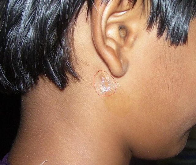
Photo 1. Pre-operative picture of parotid fistula with leakage of serous fluid from the fistulous tract and scarring of surrounding area (red circle) [1]

     Parotid fistula may be of two types

    1. Glandular: It arises directly from gland. It shows minimal discharge during rest or eating.
2. Ductal: It arises from duct. It shows profuse discharge during eating.

Parotid fistula may be extra oral or intraoral.

Extraoral fistulas are seen in the preauricular region or near the angle of mandible (see photo 1 and 2).

Photo 2. showing discharge of serous fluid from the right cheek in the angle of mandible region [2]


Causes
1. After superficial parotidectomy.
2. After drainage of parotid abscess.
3. After biopsy or Trauma.
4. Post surgical

Clinical Features
1.  Discharging fistula in the parotid region of the face, and discharge is more during eating.
2. Tenderness and induration.
3. Trismus if it gets infected

     Diagnosis
1.  Sialography to find out the origin of the fistula whether from the parotid gland or duct or ductules.
2. Fistulogram or CT fistulogram.
3. Culture of discharge if infection is suspected
4. MRI to assess soft tissues involvement

    Treatment
Ø Surgical stripping of the fistula tract
Ø Anticholinergics in post-operative period- Hyoscine bromide (Probanthine) reduce discharge
Ø Immediate post surgical fistulas can close spontaneously in such cases
Ø Newman Seabrock's operation: used for removal of anomalous arotid fistula
Ø If there is stenosis at the orifice of the Stenson's duct, papillotomy at the orifice may help.
Ø Total conservative parotidectomy is done in failed cases conserving the facial nerve
 
Ref:

Injections Techniques

Darsogluteal Intramuscular Injections


Kaposi sarcoma


Kaposi sarcoma (in AIDS):

Important points to remember about Kaposi Sarcoma

Kaposi's sarcoma is a type of cancer that forms in the lining of blood and lymph vessels.

Kaposi's sarcoma or oral cavity

Kaposi's sarcoma of the skin


Clinical Features

  • It is the most common malignancy in AIDS.
  • It is associated with the infection with a virus called the Kaposi sarcoma associated herpesvirus (KSHV), also known as human herpesvirus 8 (HHV8).
  • The commonly affected sites are palate, gingiva, tongue, and oropharynx or the skin of the face and feet.
  • It is seen early in the course of the disease. It can sometimes be confused with Bacillary angiomatosis.
  • There is a specific histologic stain for Kaposi sarcoma known as Warthin-Starry stain.
  • With the use of HAART, incidence of KS is decreasing and soon NHL may become the most common malignancy associated with AIDS.


Cemento Osseous Dysplasia

Cemento-osseous dysplasia (COD) is a benign fibro-osseous lesion of bone characterized by the replacement of normal bone by fibrous tissue and subsequently followed by its calcification with osseous and cementum-like material. It arises from the fibroblasts of the periodontal ligaments.

It is mostly asymptomatic in nature and requires no treatment. When secondarily infected, it becomes symptomatic and intervention is required.
Orthopantamogram showing a well-defined radiopaque mass in the right mandible region extending from the distal root of 45 to the mesial root of 47 [1]


As per WHO, there are three clinical presentation of cemento-osseous dysplasia.

  1. Periapical
  2. Focal
  3. Florid

Periapical cemento-osseous dysplasia

These occur in the anterior mandible and involve only a few adjacent teeth.

Focal cemento-osseous dysplasia.

involve few teeth in posterior mandible

Florid cemento-osseous dysplasia or Familial gigantiform cementoma

It is a more extensive form. it occurs bilaterally in mandible or in all jaw quadrants.
Ref:

Periapical Granuloma

The cells of periapical granuloma which are predominantly lymphocytes increase by division at the periphery. 

There are hyperaemia and oedema of the PDL; localised increase in the vascularity leads to local bone resorption mediated by osteoclast mediated delayed hypersensitivity. In the specimen slide, cholesterol crystals having needle-like appearance, and eosinic hyaline bodies known as Rushton bodies are seen. Macrophages and multinucleated giant cells are also seen. Epithelium is present.

The cells in the centre are separated from their source of nutrition; hence degenerate and liquefy. This results in an epithelium lined cavity filled with fluid known as periapical cyst.

Treatment involves RCT with apicoectomy or extraction with curettage.

Sequelae of Infection of Dental Pulp

Periapical infection with Streptococci & Staphylococci

Majority of streptococci produce hyaluronidase, an enzyme that dissolves hyaluronic acid which is a universal intercellular cementing substance. It helps in the spread of infection. Usually staphylococci are good producers of hyaluronidase, so there is no spread of infection and the infection becomes localised in the form of abscess in case of staph infection.

Oral Cancer

Oral cancer is associated with significant morbidity and mortality. Early presentations of oral cancer are usually asymptomatic, whereas late presentations include pain, discomfort, reduced mobility of the tongue, increased mobility of the teeth or an inability to wear dentures. Oral cancer varies in appearance and can mimic many other oral mucosal diseases.

Squamous cell carcinoma of the left anterior ventral surface of the tongue
Squamous cell carcinoma of the left mandibular alveolus

Oral cancer can mimic many other oral mucosal diseases, so early specialist referral is required for investigation and biopsy of any suspicious lesion. 

Any suspicious lesion needs early specialist referral for investigation and biopsy.

Squamous cell carcinoma is the most common oral malignancy, which arises from the epithelium of the oral cavity. Oral squamous cell carcinoma can affect any part of the oral mucosa; however, it most commonly occurs on the lateral surfaces of the tongue, the floor of the mouth or the gingivae. 


Risk factors for oral squamous cell carcinoma  

  1. advanced age 
  2. male gender 
  3. smoking or tobacco use 
  4. alcohol use 
  5. infection by oncogenic viruses (eg human papillomavirus) 
  6. personal or family history of squamous cell carcinoma of the head and neck 
  7. history of cancer therapy 
  8. prolonged immunosuppression 
  9. areca nut (betel quid) chewing. 
  10. Genetic susceptibility, environment, occupation and diet may also contribute to the development of oral squamous cell carcinoma. 

Cancers originating from the salivary glands and supporting nonepithelial tissues are less common than squamous cell carcinoma. Metastatic cancers to the oral soft tissues and jawbones commonly originate from primary malignancies in the breast, prostate, kidneys or lungs. Leukaemia and lymphoma may also present in the oral cavity. 


The treating specialist should perform the biopsy of an oral mucosal lesion. In rural or remote areas where a delay in specialist review is expected, seek expert advice on biopsy technique. A punch biopsy is not appropriate.


References:

  1. Therapeutic guidelines (Oral & Dental) 2019 

Oral Leukoplakia

Oral leukoplakia (OL) is a clinical term for a nonremovable white lesion that is not easily recognisable as any particular condition and therefore requires further investigation.

Oral leukoplakia manifests as patches that are bright white and sharply defined. The surfaces of the patches are slightly raised above the surrounding mucosa.

Oral leukoplakia may be homogenous (uniform lesion often with a fissured surface), or nonhomogeneous (with surface irregularity and textural or colour variation for example speckled-see below given photograph.

Stages and Phases of Anaesthesia

Assessment of the depth of anaesthesia 

Anaesthesia has been described as a series of four Stages. 

Stage 1

The period between administration of an anaesthetic and loss of consciousness. 

Stage 2

The period after loss of consciousness, which may include actions such as uncontrolled movement, delirium, vocalization.

Stage 3 

The level at which surgery can be performed. Stage 3 anaesthesia is divided into four planes

  1. Plane 1: "light" anaesthesia - the animal still has blink and swallowing reflexes, and regular respiration.
  2. Plane 2: "surgical" anaesthesia - the animal has lost blink reflexes, pupils become fixed and respiration is regular.
  3. Plane 3: "deep" anaesthesia - the animal starts losing the ability to use the respiratory muscles and breathing becomes shallow; may require assisted ventilation. The surgeries are performed in this plane.
  4. Plane 4: the animal loses all respiratory effort, and breathing may stop entirely. 

Stage 4

Anaesthetic crisis! Respiratory arrest and death from circulatory collapse imminent.

Pulpitis

Photograph showing Pulpal hyperaemia. While bacteria are still some distance from the pulp, acid permeating along the dentinal tubules gives rise to dilation of the blood vessels, oedema and a light cellular inflammatory infiltrate in the pulp [1]

What is Pulpitis?

Pulpitis is the inflammation of the pulp. It is the most common cause of pain in young persons.


Types

It is of two types.

  1. Reversible

  2. Irreversible

Irreversible pulpitis has been divided into further two types

  1. Acute pulpitis

  2. Chronic pulpitis

Causes of Pulpitis

Root Apex-Anatomical And Physiological Foramen

Anatomical and physiological root apex
Anatomical and physiological forament of the root 

Apical constriction (Physiological foramen)

  • is conside­­red narrowest diameter of the root canal

  • located at the cement-dentinal junction.

  • it is the apical limit of root canal preparation preparation.

  • it is also known as the histological foramen, because it is located at the junction between the

  • pulpal connective and interstitial loose connective tissues of the periodontal ligament.

  • not visible on x-ray (we may find it only using WL determination methods).

Dental Caries-Part 6: Prevention

Fig1: Caries free beautiful teeth with proper maintenance #

The carious process can be termed as metabolic activity in the plaque. The result of the metabolic activity may not be anything to see or there may be demineralisation resulting in a visible carious lesion. As we have discussed earlier, plaque is the cause of caries and a tooth completely free of plaque will not have caries. However, it is not always possible to show a strong association between the presence of dental plaque and caries. Mostly because people are unable to completely remove plaque themselves, even when guided properly. In addition, although the bacterial biofilm is the cause of caries, there are other factors involved. This is the reason that caries is described as a multifactorial disease. These factors, when combined, may increase or decrease the rate of demineralization. 

Dental Caries-Part 5: Clinical Aspect of Caries Pathology

Arrested caries and remineralization  


Pre-Cavitation, or “white spot” caries lesions, can stop when the balance between demineralization and remineralization is changed in favor of remineralization. This could follow the restriction of sucrose, the application of fluoride, or the loss of an adjacent tooth to a proximal caries. This loss of tooth uncovers the area of stagnation and allows proper oral hygiene procedures. The source of the calcium and phosphate for remineralization of the lesion is saliva and plaque. The caries progresses slowly, and even under natural conditions, about 50% of proximal enamel lesions may show no radiographic evidence of progression for 3 years, showing that a small change may be needed to encourage reversal of the process. Although remineralization can bring the mineral content of an enamel lesion closer to that of the original enamel, the deposition is irregular and disorganized at the single crystal level, and the structure of the original enamel cannot be recovered. Despite this, remineralized lesions that have incorporated fluoride may be less prone to caries attack than intact enamel. Arrested enamel cavities can remain dull and white or more often discolored due to the incorporation of an extrinsic dye. This is known as inactive lesion or brown spot.   

Dental Caries-Part 4: Dentine Caries

Normal Structure of Dentin


Dentine is the part of the tooth that is covered by enamel and covers the pulp tissues. It comprises of inorganic hydroxyapatite 45-50%, organic matrix 30% and water 25%.
It is a vital tissue as opposed to enamel. It is elastic and permeable. Therefore, it is never possible for a restorative material to create a totally hermetic seal between itself and the cavity wall.
Dentine consists of dentinal tubules, peritubular dentine and inter tubular dentine. Dentinal tubules contain extensions of the odontoblast cells that line along the inner surface of the dentine and remain in the pulp. The peritubular dentine makes the walls of the dentinal tubules, are devoid of collagen matrix and are highly mineralised. The inter tubular dentine is the main structural component; present between the dentinal tubules; consists of hydroxyapatite crystals lying in the collagen matrix. 

Dental Caries-Part 3: Enamel Caries


Clinical and Histological Features of Dental Caries

Now we shall describes the clinical features of carious lesions on smooth,

occlusal, and root surfaces.  We shall relate the clinical features to their histological

features. We shall consider Enamel and Dentine together, the reasons being:

  1. As a clinician, you will see them in the same way.

  2. You can not understand changes in dentine during caries progression and caries arrest without considering the spread of the enamel lesion.

  3. Changes in Dentine occur before the enamel lesion cavitates. Removal of the biofilm will arrest the lesion in dentine as well as the lesion in enamel.

  4. The lesion, in both enamel and dentine, entirely reflects the activity of the bacterial biofilm.


Before I start talking about the clinical and histological features of dental caries, You must know the 

Basic Structure of Enamel


Sound enamel consists of crystals of hydroxyapatite packed tightly together in an orderly arrangement which is known as enamel prisms. The amount of hydroxyapatite ranges between 86 to 95%; the organic component between 1% to 2 % and water between 4% to 12% by volume. The total inorganic content of enamel ranges between 95% to 98% by mass, thats why it looks like crystals.


The crystals are so tightly packed that the enamel gets a glass-like appearance and appears translucent. This is the reason that it allows the varying degrees of yellow colour of the dentine to shine through it. Here, you should know that even though the crystal packing is very tight, each crystal is actually separated from its neighbours by tiny intercrystalline spaces or pores. These spaces are filled with water and organic material. When enamel is exposed to acids produced in the microbial biofilm, mineral is removed from the surface of the crystal which shrinks in size. Thus, the intercrystalline spaces enlarge and the tissue becomes more porous. This increase in porosity can be seen clinically as a white spot.

Dental Caries-Part 3: Video series


Lecture Series on Dental Caries

To watch video lectures, click at the following links

1. Introduction

2. Etiology of dental caries

3. Bacterial plaque in dental caries

4. Microbiology of dental caries

5. Sucrose and dental caries

6. Acid production in dental plaque

7. Saliva and dental caries

8. Early enamel caries


Dental Caries-Part 2: Treatment Planning

In this chapter, you will know about the treatment planning of dental caries. In previous chapter you studies the etio-pathogenesis and clinical characteristics of dental caries. If you have not gone through it, it is advised that you study the previous chapter before proceeding further.

Mechanism of Remineralisation of Enamel

When the oral environment of a person is favourable where the pH is above 5.5 and saliva contains enough calcium and phosphate ions, the remineralisation process of enamel occurs. The supersaturated saliva acts as driving force for remineralisation. In a non cavitated enamel caries lesion, the original crystalline structure of rods remains intact. When it is etched, it acts as nucleating agent for remineralisation. When trace amount of fluoride ions is added to the environment, it enhances the remineralisation process by enhancing the precipitation of calcium and phosphate. The inclusion of fluoride ions results in the formation of fluorapatite crystals in enamel rods which is more resistant to acid attack compared to calcium apatite of the natural enamel rods. Thus, the new enamel becomes resistant to caries process. 

Dental Caries-Part 1: Etio-pathogenesis and Clinical Features

This lesson presents basic definitions, terminologies, etiologies, demineralisation-remineralisation of enamel and clinical characteristics of the caries lesion in the context of clinical operative dentistry. 

Definitions of Dental Caries and Dental Plaque 

Dental caries is defined as a multifactorial, transmissible, infectious oral disease caused primarily by the complex interaction of cariogenic oral flora (biofilm) with fermentable dietary carbohydrates on the tooth surface over time.

Dental plaque is a gelatinous mass of bacteria adhering to the tooth surface. Carious lesions occur only under the plaque. The plaque bacteria metabolises the refined carbohydrate (sucrose mainly) for energy production and produces organic acids as a by product. These acids cause dissolution of crystalline structures of enamel that result in caries lesions of the tooth.